

Motion correction in magnetic resonance spectroscopy
- PMID: 32301174
- PMCID: PMC8386494
- DOI: 10.1002/mrm.28287
Motion correction in magnetic resonance spectroscopy
Abstract
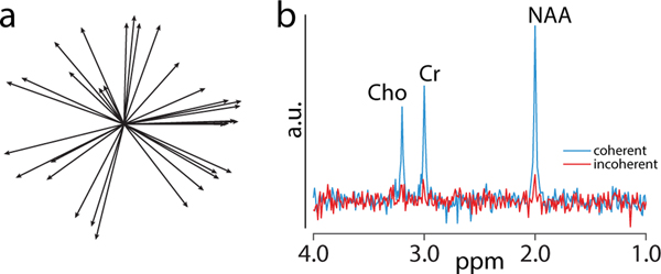
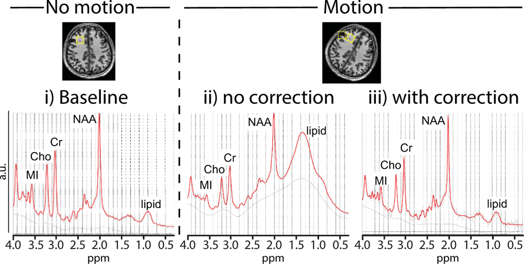
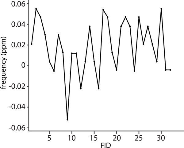
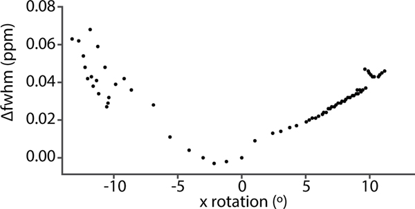
In vivo proton magnetic resonance spectroscopy and spectroscopic imaging (MRS/MRSI) are valuable tools to study normal and abnormal human brain physiology. However, they are sensitive to motion, due to strong crusher gradients, long acquisition times, reliance on high magnetic field homogeneity, and particular acquisition methods such as spectral editing. The effects of motion include incorrect spatial localization, phase fluctuations, incoherent averaging, line broadening, and ultimately quantitation errors. Several retrospective methods have been proposed to correct motion-related artifacts. Recent advances in hardware also allow prospective (real-time) correction of the effects of motion, including adjusting voxel location, center frequency, and magnetic field homogeneity. This article reviews prospective and retrospective methods available in the literature and their implications for clinical MRS/MRSI. In combination, these methods can attenuate or eliminate most motion-related artifacts and facilitate the acquisition of high-quality data in the clinical research setting.
Keywords: MRS; MRSI; motion; navigated spectroscopy sequence; prospective correction; retrospective correction.
© 2020 International Society for Magnetic Resonance in Medicine.
Figures
References
-
- De Graaf RA. In vivo NMR spectroscopy: principles and techniques: John Wiley & Sons; 2019.
-
- Teunissen C, Iacobaeus E, Khademi M, Brundin L, Norgren N, Koel-Simmelink M, Schepens M, Bouwman F, Twaalfhoven H, Blom H. Combination of CSF N-acetylaspartate and neurofilaments in multiple sclerosis. Neurology. 2009;72:1322–1329. - PubMed
-
- Schmitz B, Wang X, Barker PB, Pilatus U, Bronzlik P, Dadak M, Kahl KG, Lanfermann H, Ding XQ. Effects of aging on the human brain: a proton and phosphorus MR spectroscopy study at 3T. Journal of Neuroimaging. 2018;28:416–421. - PubMed
Publication types
MeSH terms
Grants and funding
LinkOut - more resources
Full Text Sources
Medical
