

Comment
doi: 10.7554/eLife.57105.
Rapid response
Affiliations
- PMID: 32301437
- PMCID: PMC7164951
- DOI: 10.7554/eLife.57105
Item in Clipboard
Comment
Rapid response
Elife.
.
Abstract
Extremely short X-ray pulses from a free-electron laser are helping to clarify how phytochromes respond to light, but puzzles remain.
Keywords: Deinococcus radiodurans; SFX; free-electron laser; initial photoresponse; molecular biophysics; phytochromes; structural biology.
© 2020, Hughes.
Conflict of interest statement
JH No competing interests declared
Figures
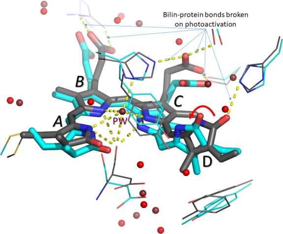
The four rings of the bilin are labelled A-D. Carbon atoms before and one picosecond after photoactivation are shown in grey and cyan, respectively. Water molecules before and after photoactivation are shown in deep red and bright red, respectively. Otherwise, oxygen, nitrogen and sulphur atoms are shown in red, blue and yellow, respectively. The ~50° rotation of the D-ring is indicated by the red arrow. The pyrrole water molecule (PW) above the nitrogen atoms of the A-, B- and C-rings disappears on photoactivation. Hydrogen bonds (yellow dashes) between the bilin and amino acid side chains in the rest of the phytochrome are also broken. Figure prepared by the author using PyMol from data provided by Claesson et al.
Comment on
-
The primary structural photoresponse of phytochrome proteins captured by a femtosecond X-ray laser.Elife. 2020 Mar 31;9:e53514. doi: 10.7554/eLife.53514. Elife. 2020. PMID: 32228856 Free PMC article.
References
-
- Claesson E, Wahlgren WY, Takala H, Pandey S, Castillon L, Kuznetsova V, Henry L, Panman M, Carrillo M, Kübel J, Nanekar R, Isaksson L, Nimmrich A, Cellini A, Morozov D, Maj M, Kurttila M, Bosman R, Nango E, Tanaka R, Tanaka T, Fangjia L, Iwata S, Owada S, Moffat K, Groenhof G, Stojković EA, Ihalainen JA, Schmidt M, Westenhoff S. The primary structural photoresponse of phytochrome proteins captured by a femtosecond X-ray laser. eLife. 2020;9:e53514. doi: 10.7554/eLife.53514. - DOI - PMC - PubMed
Publication types
MeSH terms
Substances
LinkOut - more resources
Full Text Sources
