. 2020 Apr 17;15(4):e0231423.
doi: 10.1371/journal.pone.0231423. eCollection 2020.

Recapitulation of the accessible interface of biopsy-derived canine intestinal organoids to study epithelial-luminal interactions

Affiliations

Recapitulation of the accessible interface of biopsy-derived canine intestinal organoids to study epithelial-luminal interactions

Yoko M Ambrosini et al. PLoS One. .

Abstract

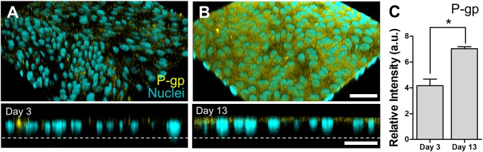
Recent advances in canine intestinal organoids have expanded the option for building a better in vitro model to investigate translational science of intestinal physiology and pathology between humans and animals. However, the three-dimensional geometry and the enclosed lumen of canine intestinal organoids considerably hinder the access to the apical side of epithelium for investigating the nutrient and drug absorption, host-microbiome crosstalk, and pharmaceutical toxicity testing. Thus, the creation of a polarized epithelial interface accessible from apical or basolateral side is critical. Here, we demonstrated the generation of an intestinal epithelial monolayer using canine biopsy-derived colonic organoids (colonoids). We optimized the culture condition to form an intact monolayer of the canine colonic epithelium on a nanoporous membrane insert using the canine colonoids over 14 days. Transmission and scanning electron microscopy revealed a physiological brush border interface covered by the microvilli with glycocalyx, as well as the presence of mucin granules, tight junctions, and desmosomes. The population of stem cells as well as differentiated lineage-dependent epithelial cells were verified by immunofluorescence staining and RNA in situ hybridization. The polarized expression of P-glycoprotein efflux pump was confirmed at the apical membrane. Also, the epithelial monolayer formed tight- and adherence-junctional barrier within 4 days, where the transepithelial electrical resistance and apparent permeability were inversely correlated. Hence, we verified the stable creation, maintenance, differentiation, and physiological function of a canine intestinal epithelial barrier, which can be useful for pharmaceutical and biomedical researches.


Conflict of interest statement

J.P.M., A.E.J., K.A., and H.J.K. are co-founders of 3D Health Solutions Inc. and hold an equity interest in the company. T.A. is employed by and receives a salary from 3D Health Solutions Inc. This does not alter our adherence to PLOS ONE policies on sharing data and materials.

Figures

Fig 1
Fig 1. Morphological analysis of the 3D colonoids and the 2D canine colonic monolayer.
(A) A growth profile of the colonoid isolated from the canine colonic crypt. A small spherical colonoids progressively grows to form fully grown colonoids. Representative phase-contrast micrographs were taken at days 1, 3, and 5. The zoomed-in inset at each day shows the high-power magnification of a colonoid in the white dashed box. (B) A schematic displays the procedure of the formation of an epithelial monolayer derived from 3D canine colonoids. The fully-grown organoids are dissociated into single cells, then seeded into a nanoporous insert to form a monolayer. AP, apical; BL, basolateral. (C) Representative phase-contrast micrographs on day 3 and 13 are provided, respectively. Bars, 200 μm.
Fig 2
Fig 2. Electron microscopic characterization of the apical surface and the tissue interface of the canine colonoid-derived monolayer.
(A) A low magnification SEM image of the microvilli on the apical cell surface. (B) A high-power magnification of the microvilli from A indicated by a white dashed box. Bars, 5 μm. (C) A TEM image of the microvilli on the cell monolayer. MV, microvilli. Bar, 500 nm. (D) A high-power TEM image that shows the microvilli (MV) and the surrounding glycocalyx (GLX). Bar, 200 nm.
Fig 3
Fig 3. The cell type-specific characterization of the canine colonoid-derived epithelial monolayer.
The canine colonoid-derived monolayer on Day 13 was used to visualize the markers highlighting the cell lineages, proliferation, and mucus production. The population of stem cells (A; Lgr5+, Yellow), proliferative cells (B; Ki67, Red), absorptive enterocytes (C; ALPI, Magenta), and enteroendocrine cells (D; Neurog3, Red and E; CgA, Red) were visualized by using RNA in situ hybridization (for A, C, and D) or IF staining (for B and E). As a counterstaining, E-cadherin (Cyan for A, C, and D), F-actin (Green for B and Cyan for E), or nuclei (Grey for A, B, C, D, and E) were displayed. Bars, 20 μm. (F) Quantification of the population of the cells that show positive signals to the target markers normalized by the total numbers of nuclei. Three independent fields of view from two or more independent biological replicates were used. In each biological replicate, 2 technical replicates were performed. Error bars indicate SEM.
Fig 4
Fig 4. Visualization of mucus production and goblet cells in the canine colonoid-derived monolayer.
The mucus production (A; WGA) was visualized by live-cell imaging at the apical surface of the monolayer. Bar, 20 μm. (B) A representative TEM image shows the goblet cell with multiple mucin granules (MG) and mitochondria (M). Bar, 1 μm. (C) A low magnification SEM image of a goblet cell on the apical cell surface of the canine colonoid-derived monolayer. Bar, 5 μm. (D) A high magnification of a goblet cell orifice (GO), a fenestrated membrane (FM) extending deep into the cell, and microvilli (MV) from C indicated by a white dashed box. Bar, 1 μm.
Fig 5
Fig 5. Expression of the P-gp in the canine colonoid-derived monolayer.
The expression of P-gp was visually characterized by IF staining. Angled (upper) and cross-sectional side views (lower) show the localization the P-gp proteins (Yellow) on the polarized colonoid-derived monolayer at days 3 (A) and 13 (B), respectively. Nuclei, Cyan. Dashed lines pinpoint the location of the basement membrane in the nanoporous insert. Bars, 50 μm. (C) Quantification of the P-gp expression at days 3 and 13, respectively. Total 10 randomly chosen fields of view were used to detect P-gp expression levels among 4 biological replicates. In each biological replicate, we performed 2 technical replicates. a.u., arbitrary unit. Error bars indicate SEM.*P<0.0001.
Fig 6
Fig 6. Characterization of the junctional proteins and the barrier function in the canine colonoid-derived epithelial monolayer.
Visualization of the spatial localization of the ZO-1 (A; Magenta) and E-cadherin (B; Cyan) on the same location of a canine colonoid-derived monolayer. Nuclei, Grey. Bar, 50 μm. (C) The profile of the epithelial barrier function was monitored by measuring TEER. The effect of culture medium on TEER was demonstrated by applying the regular proliferation medium in both the apical and basolateral side of the Transwell (Control, open circle) versus the differentiation/proliferation medium in the apical/basolateral compartments, respectively (Diff; closed circle). Both groups were cultured with the proliferation medium by Day 4 (a dashed line), then different culture media were applied (Diff vs. Control) for additional 4 days. Two biological replicates with 4 technical replicates were used in each condition. *P<0.01. (D) A TEM image of the intercellular junctional complex in the canine colonoid-derived monolayer and a zoom-in (E) that shows a high-power magnification of the white dashed area in D. MV, microvilli; M, mitochondria; and D, desmosome. Bars, 500 nm. (F) The profile of TEER (open circle) and corresponding apparent permeability (Papp) of fluorescein (closed square) on the days of 2, 3, 5, and 6 of the cultures. Each data point was prepared with 2 biological and 4 technical replicates. Error bars indicate SEM.

References

    1. Kol A, Arzi B, Athanasiou KA, Farmer DL, Nolta JA, Rebhun RB, et al. Companion animals: Translational scientist’s new best friends. Sci Transl Med. 2015. October 7;7(308):308ps21 10.1126/scitranslmed.aaa9116 - DOI - PMC - PubMed
    1. Schneider B, Balbas‐Martinez V, Jergens AE, Troconiz IF, Allenspach K, Mochel JP. Model‐Based Reverse Translation Between Veterinary and Human Medicine: The One Health Initiative. CPT Pharmacomet Syst Pharmacol. 2018. February;7(2):65–8. - PMC - PubMed
    1. Axelsson E, Ratnakumar A, Arendt M-L, Maqbool K, Webster MT, Perloski M, et al. The genomic signature of dog domestication reveals adaptation to a starch-rich diet. Nature. 2013. March 21;495(7441):360–4. 10.1038/nature11837 - DOI - PubMed
    1. Coelho LP, Kultima JR, Costea PI, Fournier C, Pan Y, Czarnecki-Maulden G, et al. Similarity of the dog and human gut microbiomes in gene content and response to diet. Microbiome. 2018. April 19;6(1):72 10.1186/s40168-018-0450-3 - DOI - PMC - PubMed
    1. Gartzke D, Fricker G. Establishment of optimized MDCK cell lines for reliable efflux transport studies. J Pharm Sci. 2014. April;103(4):1298–304. 10.1002/jps.23901 - DOI - PubMed

Publication types

Substances