. 2020 Jun 4;26(6):880-895.e6.
doi: 10.1016/j.stem.2020.03.013. Epub 2020 Apr 16.

Dermal Adipocyte Lipolysis and Myofibroblast Conversion Are Required for Efficient Skin Repair

Affiliations

Dermal Adipocyte Lipolysis and Myofibroblast Conversion Are Required for Efficient Skin Repair

Brett A Shook et al. Cell Stem Cell. .

Abstract

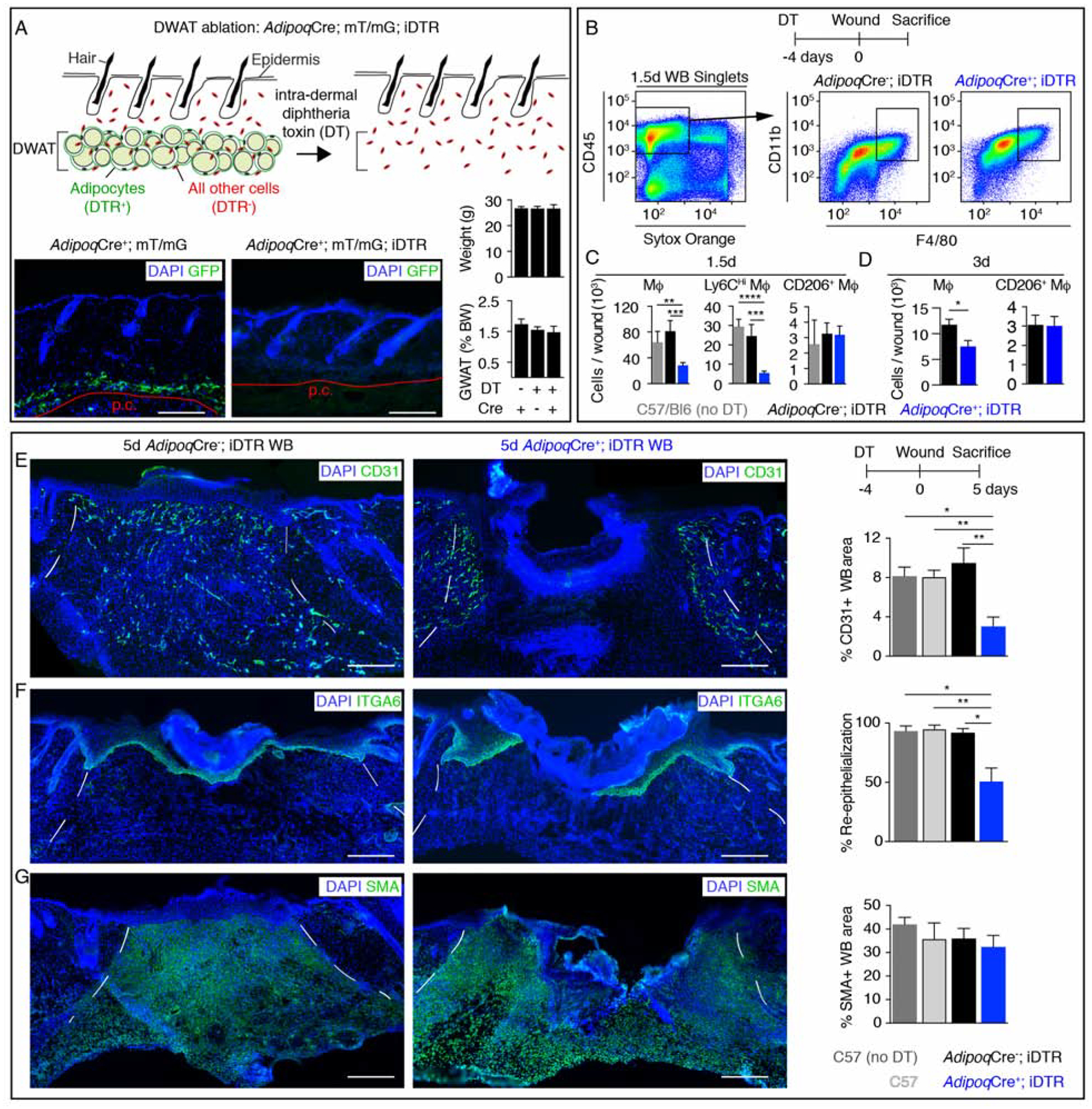
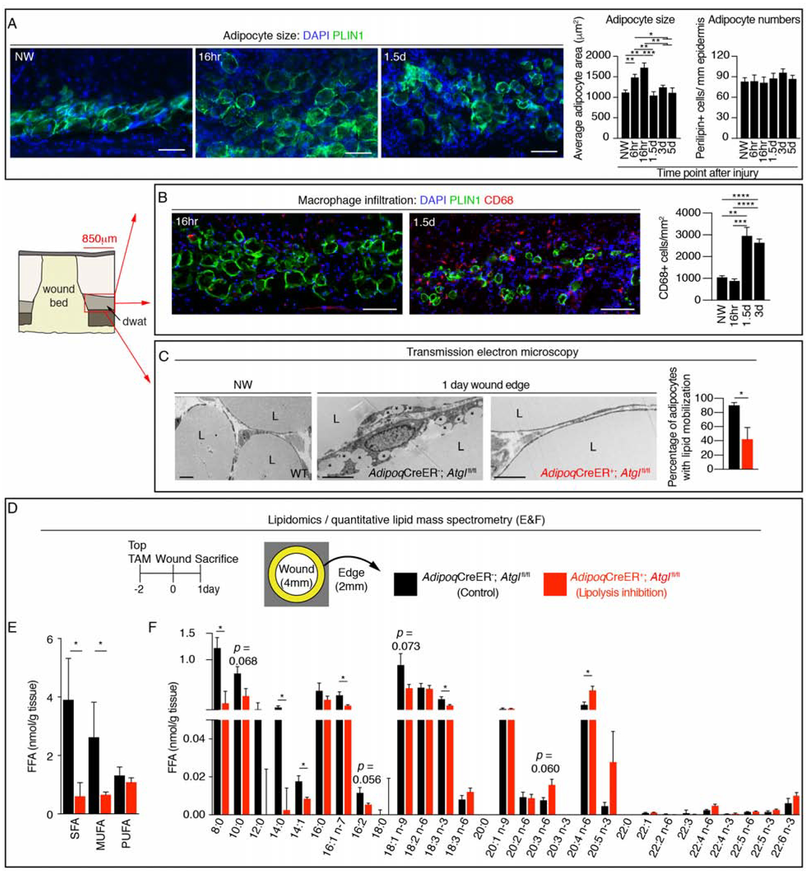
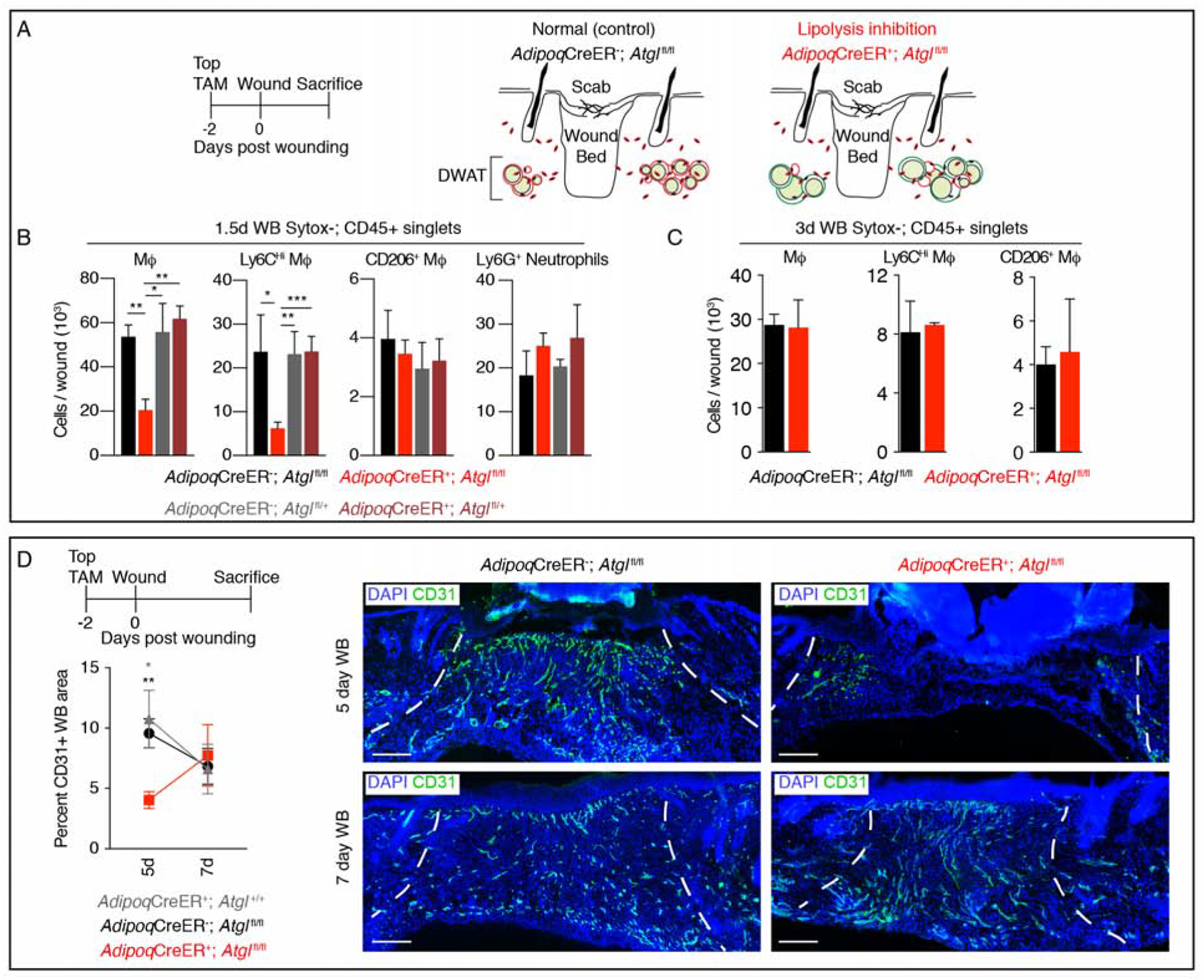
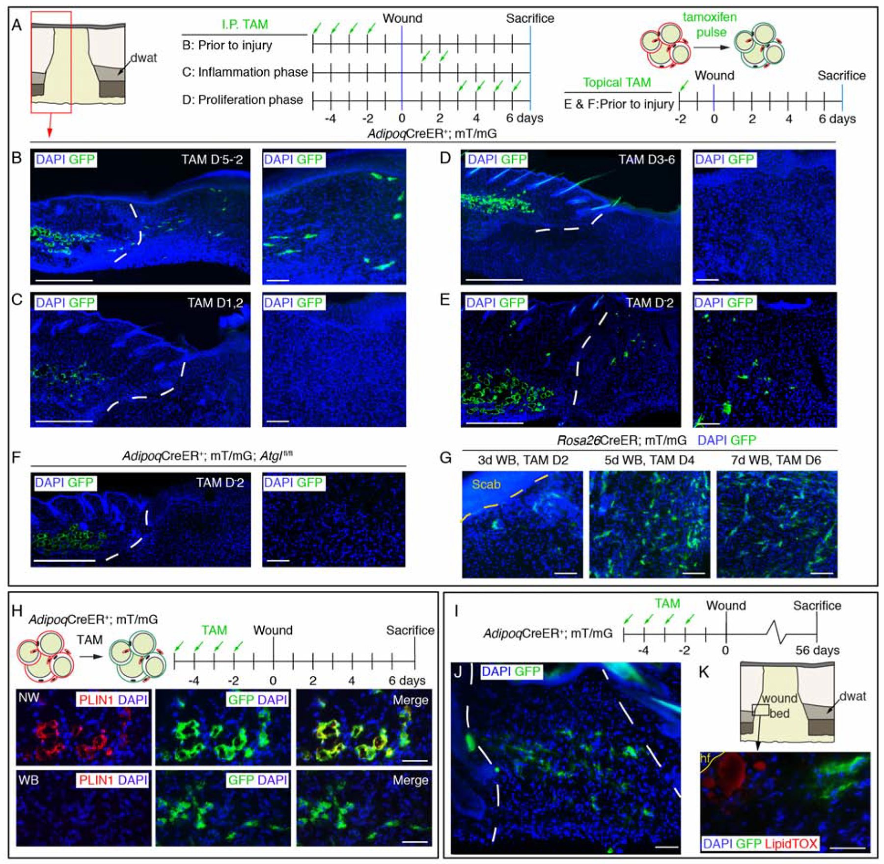
Mature adipocytes store fatty acids and are a common component of tissue stroma. Adipocyte function in regulating bone marrow, skin, muscle, and mammary gland biology is emerging, but the role of adipocyte-derived lipids in tissue homeostasis and repair is poorly understood. Here, we identify an essential role for adipocyte lipolysis in regulating inflammation and repair after injury in skin. Genetic mouse studies revealed that dermal adipocytes are necessary to initiate inflammation after injury and promote subsequent repair. We find through histological, ultrastructural, lipidomic, and genetic experiments in mice that adipocytes adjacent to skin injury initiate lipid release necessary for macrophage inflammation. Tamoxifen-inducible genetic lineage tracing of mature adipocytes and single-cell RNA sequencing revealed that dermal adipocytes alter their fate and generate ECM-producing myofibroblasts within wounds. Thus, adipocytes regulate multiple aspects of repair and may be therapeutic for inflammatory diseases and defective wound healing associated with aging and diabetes.

Keywords: cellular plasticity; dermal adipose tissue; inflammation; lipolysis; myofibroblast; wound healing.


Conflict of interest statement

Declaration of Interests The authors have no competing interests.

Figures

Figure 1.
Figure 1.. Dermal adipocytes contribute to skin wound healing.
(A) Schematic and GFP immunostained images of uninjured skin and quantification of mouse body weight (BW) and the percentage of BW that is gonadal white adipose tissue (GWAT), following intradermal injection of diphtheria toxin (n ≥ 6 mice for each condition). Red line delineates the panniculus carnosus (p.c.). Scale bars, 250μm. (B–D) Flow cytometry plots (B) and quantification (C–D) of macrophages after injury (n ≥ 5 mice each condition and time point). (E–G) Immunostained sections and quantification of CD31 (revascularization) (E) (n ≥ 6 mice each condition), ITGA6 (re-epithelialization) (F) (n ≥ 6 mice each condition) and smooth muscle actin (SMA; fibroblast repopulation) (G) (n ≥ 4 mice each condition) in day 5 wound beds (WB). Scale bars, 250μm. Dotted white lines delineate wound edges. Error bars indicate mean ± SEM. *, p < 0.05; **, p < 0.01; ***, p < 0.001, ****, p < 0.0001. DT, diphtheria toxin. See also Figure S1 and S2.
Figure 2.
Figure 2.. Dermal adipocytes undergo lipolysis after injury.
(A) Representative PLIN1 immunostained images of DWAT at the periphery of wounds and quantification of adipocyte cross sectional area and numbers (n ≥ 6 mice each time point). Scale bars, 100μm. (B) Representative immunostained images and quantification of CD68+ cells at the periphery of wound beds and in non-wounded (NW) skin (n ≥ 6 mice each time point). Scale bars, 100μm. (C) Transmission electron microscopy of adipocytes in uninjured skin and adipocytes at the wound periphery 1 day after injury. Asterisks show small lipid droplets. Quantification of adipocytes containing small lipid droplets at the periphery of a larger lipid droplet (n = 4 mice each condition). Scale bars, 5μm. (D) Experimental approach for quantitative lipidomics. (E–F) Lipid mass spectrometry quantification of non-esterified free fatty acid (FFA) classes (E) (n = 4–5 mice each condition) and FFA species (F) (n = 4–5 mice each time point). L, lipid droplet; SFA, saturated fatty acid; MUFA, monounsaturated fatty acid; PUFA, polyunsaturated fatty acid. Error bars indicate mean ± SEM. *, p < 0.05; **, p < 0.01; ***, p < 0.001, ****, p < 0.0001. See also Figure S3.
Figure 3.
Figure 3.. Impairing adipocyte lipolysis reduces macrophage numbers during inflammation.
(A) Schematic of the strategy to inhibit lipolysis during wound healing. (B) Flow cytometry quantification of macrophages, macrophage subsets and neutrophils in 1.5-day wound beds (WB) (n = 5–7 mice each condition). (C) Flow cytometry quantification of macrophages and macrophage subsets in 3-day wound beds (n = 3–4 mice each condition). (D) CD31 immunostained sections and quantification 5 and 7 days after injury (n = 6 mice 5-day; n ≥ 3 mice 7-day). Scale bars, 250μm. Dotted lines indicate wound edges. Error bars indicate mean ± SEM. *, p < 0.05; **, p < 0.01; ***, p < 0.001. See also Figure S4.
Figure 4.
Figure 4.. Adipocyte-derived cells migrate into wound beds.
(A–F) Schematic of tamoxifen labeling strategy and immunostained images of GFP+ cells in day 7 wounds. Lower magnification images show dermal adipocytes (GFP+) and wound beds. Higher magnification panels are representative images from inside wound beds. Red box in A is representative of the wound bed location displayed in low magnification images in B–F. Scale bars, 500μm in lower magnification panels and 100μm in higher magnification panels. (G) Images of GFP+ cells in the center of wound beds from Rosa26CreER; mT/mG mice treated with tamoxifen at different stages of wound healing. (H) Schematic of adipocyte lineage tracing and PLIN1 immunostained sections in non-wounded (NW) skin and inside 7-day wound beds (WB). Scale bars, 100μm. (I) Labeling scheme to identify AdipoqCreER-traced cells. (J) Images of GFP+ cells in wound beds 8 weeks after injury. (K) LipidTOX staining at the wound edge near a growing peripheral hair follicle (hf). Scale bars, 50μm. White dotted lines delineate wound edges. See also Figure S5.
Figure 5.
Figure 5.. Adipocyte-derived cells in wound beds are long-lived and do not colocalize with adipocyte markers.
(A) Images of GFP in sections from the center of wound beds at different time points after injury. Scale bars, 500μm in lower magnification and 100μm in higher magnification (designated by orange box). (B) Quantification of GFP+ cells in wound beds (n = 4 mice each time point) and (C) the spatial distribution of GFP signal (n ≥ 7 mice each time point). (D) Colocalization of PLIN1 or PPARg with GFP+ adipocytes at the periphery of wounds 16-hours after injury. Dotted box indicates an area of GFP+ cells that lack PLIN1. Arrows indicate GFP+; PPARg+ cells, arrow heads indicate GFP+; PPARg− cells. Scale bars, 100μm. (E) PLIN1 and GFP immunostained images of DWAT and quantification of regions devoid of PLIN1 (orange lines) or GFP (magenta lines) at the periphery of wound beds at different time points after injury (n ≥ 6 mice each time point). Scale bars, 100μm. (F) Labeling scheme to identify long-lived AdipoqCreER-traced cells and PPARg staining. Arrows indicate GFP+; PPARg+ cells, arrow heads indicate GFP+; PPARg− cells. Scale bars, 50μm. (G) Immunostaining of PLIN1 or PPARg with GFP in wound periphery adipocytes from AdipoqCreER+; mT/mG; Atglfl/fl mice 16 hours after injury. Arrows indicate GFP+; PLIN1+ or GFP+; PPARg+ cells, arrow heads indicate GFP+; PPARg− cells. Scale bars, 200μm. White dotted lines delineate the wound edge. A.U., arbitrary units. Error bars indicate mean ± SEM. See also Figure S5.
Figure 6.
Figure 6.. Adiponectin traced cells gain a myofibroblast gene expression profile during skin repair.
(A) Schematic and principal component (PC) analysis of cell populations isolated for RNA-seq. (B) Number of differentially expressed genes between cellular subsets. (C) Fold change in gene expression of myofibroblast-associated genes in GFP+ cells relative to wound edge/periphery adipocytes. Data from RNA-seq. (D) Quantitative real-time PCR of adipocyte signature genes (left) and myofibroblast genes (right) in GFP+ cells relative to wound periphery adipocytes (n = 3 mice). (E) Genes upregulated in GFP+ cells relative to wound edge adipocytes. Genes in red are also enriched compared to CD26+ myofibroblasts. (F–G) Images from day 5 wounds of AdipoqCre+; mT/mG mice (F) and quantification (G) of GFP+ cell colocalization with myofibroblast-associated markers (n = 3–5 mice). Scale bars, 10μm. (H) Flow cytometry plot and quantification of EdU+ cells (n = 3 mice). Scale bar, 100μm. Error bars indicate mean ± SEM. Epi, epidermis; d, dermis; dwat, dermal white adipose tissue; WB, wound bed. See also Figure S6.
Figure 7.
Figure 7.. Dermal adipocyte-derived cells become myofibroblasts after injury.
(A) scRNA-seq was performed on non-wounded (NW) skin and day 5 wound beds (WB) from AdipoqCre; mT/mG mice. UMAP dimension reduction plot is displayed with a table (B) identifying cell clusters based on enriched genes and quantifying their relative abundance. (C) Gene expression plots showing distribution of Acta2, Col1a1, Col3a1, Pdgfra and Vimentin. Expression levels for each cell are shown as Pearson residuals and displayed using a color scale, overlaid onto the UMAP plot. GFP+ cells from NW and WB samples are overlaid to emphasize their location. (D) Schematic illustration showing the contribution of adipocytes to skin wound healing. Following injury, adipocytes undergo lipolysis that supports macrophage inflammation. Adipocytes depleted of lipids become myofibroblasts and proliferate. Depletion of dermal adipocytes and inhibition of dermal adipocyte lipolysis reduces macrophage numbers during inflammation and delays revascularization. Ablating dermal adipocytes also delays re-epithelialization. FB, fibroblast; VSMC, vascular smooth muscle cell. See also Figure S7, Table S1 and Table S2.

Comment in

References

    1. Alvarez-Curto E, and Milligan G (2016). Metabolism meets immunity: The role of free fatty acid receptors in the immune system. Biochemical Pharmacology 114, 3–13. - PubMed
    1. Bani D, Li AQ, Freschi G, and Russo GL (2013). Histological and Ultrastructural Effects of Ultrasound-induced Cavitation on Human Skin Adipose Tissue. Plastic and Reconstructive Surgery Global Open 1, e41–10. - PMC - PubMed
    1. Barrientos S, Stojadinovic O, Golinko MS, Brem H, and Tomic-Canic M (2008). Growth factors and cytokines in wound healing. Wound Repair and Regeneration 16, 585–601. - PubMed
    1. Benjamini Y, Drai D, Elmer G, Kafkafi N, and Golani I (2001). Controlling the false discovery rate in behavior genetics research. Behav. Brain Res 125, 279–284. - PubMed
    1. Berndt J, Kralisch S, Klöting N, Ruschke K, Kern M, Fasshauer M, Schön M, Stumvoll M, and Blüher M (2008). Adipose Triglyceride Lipase Gene Expression in Human Visceral Obesity. Exp Clin Endocrinol Diabetes 116, 203–210. - PubMed

Publication types