Review
. 2020 Aug:64:35-42.
doi: 10.1016/j.clinimag.2020.04.001. Epub 2020 Apr 8.

Portable chest X-ray in coronavirus disease-19 (COVID-19): A pictorial review

Affiliations
Review

Portable chest X-ray in coronavirus disease-19 (COVID-19): A pictorial review

Adam Jacobi et al. Clin Imaging. 2020 Aug.

Abstract

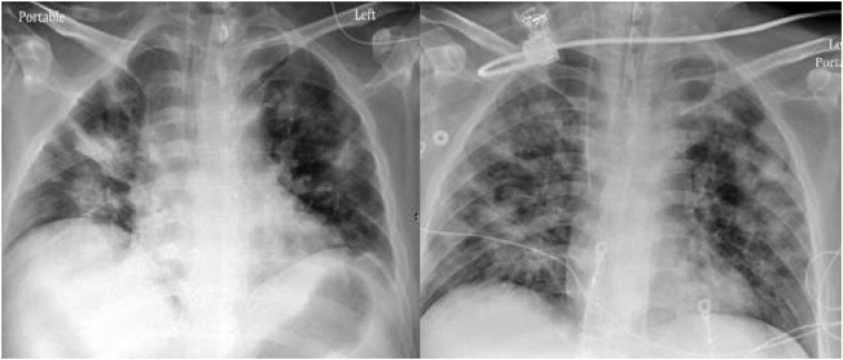
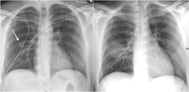
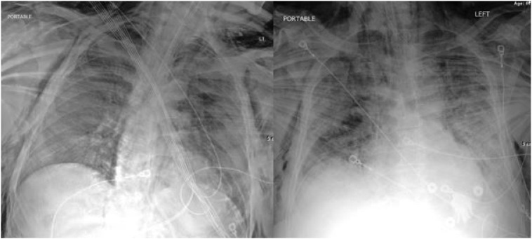
As the global pandemic of coronavirus disease-19 (COVID-19) progresses, many physicians in a wide variety of specialties continue to play pivotal roles in diagnosis and management. In radiology, much of the literature to date has focused on chest CT manifestations of COVID-19 (Zhou et al. [1]; Chung et al. [2]). However, due to infection control issues related to patient transport to CT suites, the inefficiencies introduced in CT room decontamination, and lack of CT availability in parts of the world, portable chest radiography (CXR) will likely be the most commonly utilized modality for identification and follow up of lung abnormalities. In fact, the American College of Radiology (ACR) notes that CT decontamination required after scanning COVID-19 patients may disrupt radiological service availability and suggests that portable chest radiography may be considered to minimize the risk of cross-infection (American College of Radiology [3]). Furthermore, in cases of high clinical suspicion for COVID-19, a positive CXR may obviate the need for CT. Additionally, CXR utilization for early disease detection may also play a vital role in areas around the world with limited access to reliable real-time reverse transcription polymerase chain reaction (RT-PCR) COVID testing. The purpose of this pictorial review article is to describe the most common manifestations and patterns of lung abnormality on CXR in COVID-19 in order to equip the medical community in its efforts to combat this pandemic.

Keywords: COVID-19; Chest CT; Chest X-ray; Coronavirus.


Figures

Fig. 1
Fig. 1
Portable CXR (left) with vague hazy densities in the right upper lobe (white arrow) which correspond to ground glass opacities (black arrow) on coronal image from contrast enhanced CT (right) performed the same day.
Fig. 2
Fig. 2
CXR (left) with patchy peripheral left mid to lower lung opacities (black arrow) corresponding to ground glass opacities (white arrow) on coronal image from contrast-enhanced the contemporaneous chest CT (right).
Fig. 3
Fig. 3
CXR (left) with reticular and hazy left lower lobe opacities (black arrow) in a patient with COVID-19. Similar findings are present on the coronal CT from the same day (right).
Fig. 4
Fig. 4
CXR (left) with subtle ill-defined hazy opacities in the right (black arrows) greater than left lungs in a patient with COVID-19. Findings are easier to appreciate on subsequent CT the same day (right).
Fig. 5
Fig. 5
Six different patients with varying degrees of COVID-19 pneumonia predominantly involving the lower lung zones (black arrows) bilaterally on CXR.
Fig. 6
Fig. 6
Four different patients with varying degrees of COVID-19 pneumonia on CXR predominantly involving the peripheral lungs bilaterally (black arrows).
Fig. 7
Fig. 7
CXR (left) and subsequent coronal image from chest CT (right) performed in a patient with COVID-19 and diffuse ground glass and consolidative opacities throughout both lungs.
Fig. 8
Fig. 8
Two different intubated patients with COVID-19 infection and diffuse lung opacities.
Fig. 9
Fig. 9
Serial radiographs over 7 days in a patient with COVID-19 infection depicting progression of diffuse lung disease that ultimately required intubation.
Fig. 10
Fig. 10
Serial chest radiographs of a different patient with COVID-19 infection separated by 6 days depicting progression of diffuse lung disease requiring intubation.
Fig. 11
Fig. 11
Serial chest radiographs of a COVID-19 patient with diffuse lung opacities (left image) with interval cavitation (left image white arrows) and tension pneumothorax (right image black arrow). Successful chest tube placement was subsequently performed (not shown).
Fig. 12
Fig. 12
A unique case of a rapidly enlarging right lung nodule (left image, white arrow) over the course of 5 days. Subsequent bronchoscopy with tissue sampling revealed COVID-19 infection.
Fig. 13
Fig. 13
Two different intubated patients with COVID-19 and diffuse subcutaneous emphysema and pneumomediastinum identified on CXR. No pneumothorax was identified.

References

    1. Zhou S., Wang Y., Zhu T., Xia L. CT features of coronavirus disease 2019 (COVID-19) pneumonia in 62 patients in Wuhan, China. Am J Roentgenol. 2020:1–8. March. - PubMed
    1. Chung M., Bernheim A., Mei X. CT imaging features of 2019 novel coronavirus (2019-nCoV) Radiology. February 2020;200230 doi: 10.1148/radiol.2020200230. - DOI - PMC - PubMed
    1. ACR recommendations for the use of chest radiography and computed tomography (CT) for suspected COVID-19 infection|American College of Radiology. https://www.acr.org/Advocacy-and-Economics/ACR-Position-Statements/Recom.... Accessed March 22, 2020. Google Scholar 2020.
    1. Wong H.Y.F., Lam H.Y.S., Fong A.H. Frequency and distribution of chest radiographic findings in COVID-19 positive patients. Radiology. 2019:201160. doi: 10.1148/radiol.2020201160. Mar 27. - DOI - PMC - PubMed
    1. Vilar J., Domingo M.L., Soto C. Radiology of bacterial pneumonia. J Eur J Radiol. 2004 Aug;51(2):102–113. - PubMed

Substances