. 2020 Apr 17;21(1):30.
doi: 10.1186/s12860-020-00274-1.

The mitochondrial outer membrane protein SYNJ2BP interacts with the cell adhesion molecule TMIGD1 and can recruit it to mitochondria

Affiliations

The mitochondrial outer membrane protein SYNJ2BP interacts with the cell adhesion molecule TMIGD1 and can recruit it to mitochondria

Christian Hartmann et al. BMC Mol Cell Biol. .

Abstract

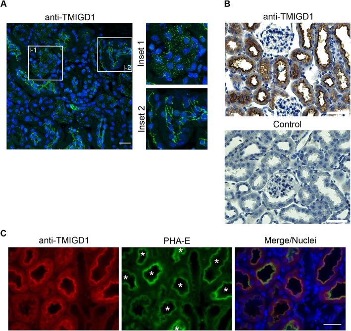
Background: Transmembrane and immunoglobulin domain-containing protein 1 (TMIGD1) is a recently identified cell adhesion molecule which is predominantly expressed by epithelial cells of the intestine and the kidney. Its expression is downregulated in both colon and renal cancer suggesting a tumor suppressive activity. The function of TMIGD1 at the cellular level is largely unclear. Published work suggests a protective role of TMIGD1 during oxidative stress in kidney epithelial cells, but the underlying molecular mechanisms are unknown.

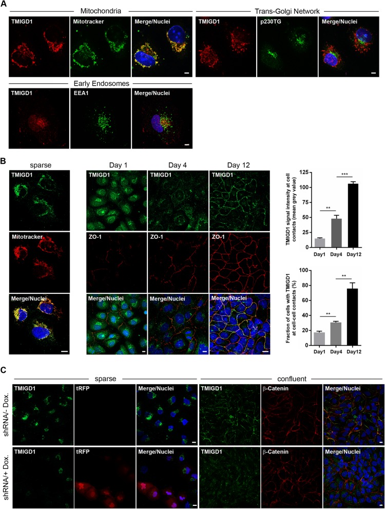
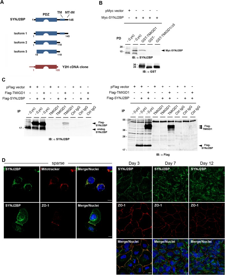
Results: In this study, we address the subcellular localization of TMIGD1 in renal epithelial cells and identify a cytoplasmic scaffold protein as interaction partner of TMIGD1. We find that TMIGD1 localizes to different compartments in renal epithelial cells and that this localization is regulated by cell confluency. Whereas it localizes to mitochondria in subconfluent cells it is localized at cell-cell contacts in confluent cells. We find that cell-cell contact localization is regulated by N-glycosylation and that both the extracellular and the cytoplasmic domain contribute to this localization. We identify Synaptojanin 2-binding protein (SYNJ2BP), a PDZ domain-containing cytoplasmic protein, which localizes to both mitochondria and the plasma membrane, as interaction partner of TMIGD1. The interaction of TMIGD1 and SYNJ2BP is mediated by the PDZ domain of SYNJ2BP and the C-terminal PDZ domain-binding motif of TMIGD1. We also find that SYNJ2BP can actively recruit TMIGD1 to mitochondria providing a potential mechanism for the localization of TMIGD1 at mitochondria.

Conclusions: This study describes TMIGD1 as an adhesion receptor that can localize to both mitochondria and cell-cell junctions in renal epithelial cells. It identifies SYNJ2BP as an interaction partner of TMIGD1 providing a potential mechanism underlying the localization of TMIGD1 at mitochondria. The study thus lays the basis for a better understanding of the molecular function of TMIGD1 during oxidative stress regulation.

Keywords: Adhesion molecule; Cell-cell adhesion; JAM; Kidney epithelium; SYNJ2BP; TMIGD1.


Conflict of interest statement

The authors declare that they have no competing interests.

Figures

Fig. 1
Fig. 1
Ectopic TMIGD1 is localized in the cytoplasm of MDCKII cells but is efficiently transported to the cell surface after deletion of its D1 domain. a Schematic presentation of Flag-tagged TMIGD1 constructs expressed in MDCKII-TetOFF cells. The two ΔD1 mutants lack the membrane-distal Ig-like domain (D1 domain). The PDZ domain-binding motif (shown in red) is absent in the “Δ5” mutants. b MDCKII cell lines stably transfected with the TMIGD1 constructs depicted in (a) (TMIGD1/WT (WT), TMIGD1/Δ5 (Δ5), ΔD1-TMIGD1 (ΔD1), ΔD1-TMIGD1/Δ5 (ΔD1−/Δ5) were induced by doxycycline removal to express the transgenes and analyzed by Western blotting with antibodies against the Flag tag. c Flag-TMIGD1-expressing MDCKII-TetOFF cells were analyzed for cell surface localization of TMIGD1 constructs by flow cytometry using antibodies against the Flag tag. Cells were left untreated (Not permeabilized) to analyze cell surface proteins only, or were permeabilized by saponin treatment (Permeabilized) to analyze total protein levels. Note that the constructs with a complete extracellular domain (TMIGD1/WT, TMIGD1/Δ5) are predominantly localized in the cytoplasm, whereas constructs lacking the membrane-distal Ig-like domain (ΔD1-TMIGD1, ΔD1-TMIGD1/Δ5) are localized at the cell surface. Filled grey lines indicate unstained cells, dotted black lines indicate cells stained with secondary antibodies alone. d Immunofluorescence analysis of TMIGD1-expressing MDCKII cell lines depicted in panel (a). Cells were stained with antibodies against the Flag tag and against β-catenin. Nuclei were stained with DAPI (pseudocoloured in white). Note that the constructs lacking the D1 Ig-like domain are localized at cell-cell contacts. Scale bars: 5 μm. e Quantification of cell-cell contact localization of TMIGD1 constructs. Cells were visually inspected for Flag signals at cell-cell contacts. TMIGD1 signals were rated as cell-cell contact-localized when a clear overlap with β-catenin signals at linear cell-cell contact sites was observed. Statistical analysis was performed using unpaired Student’s t-test. Data were obtained from three independent experiments and are presented as arithmetic means ± SEM; ****P < 0.0001. The differences in cell-cell contact localization between the two ΔD1-TMIGD1 constructs (ΔD1-TMIGD1, ΔD1-TMIGD1/Δ5) and each of the two constructs with entire extracellular domains (TMIGD1, TMIGD1/Δ5) are highly significant (P < 0.0001; not indicated by asterisks in the graph). f Cell-cell contact localization of TMIGD1 - JAM-A domain-swapping mutants. Left panel: Schematic presentation of domain swapping mutants. Middle panel: TMIGD1 - JAM-A swapping mutants shown in the left panel were transiently expressed in MDCKII cells. The subcellular localization was analyzed by fluorescence microscopy based on the EGFP signals. Right panel: Quantification of cell-cell contact localization of the TMIGD1 - JAM-A domain-swapping mutants. Cells were visually inspected for the localization of EGFP fluorescence signals at linear cell-cell contacts defined by β-catenin-positive signals (not shown). Statistical analyses were performed using unpaired Student’s t-test and display comparisons of the TMIGD1 swapping mutants with the TMIGD1 full length protein (TMIGD1-EGFP). Data were obtained from three independent experiments and are presented as arithmetic means ± SEM; ***P < 0.001
Fig. 2
Fig. 2
TMIGD1 is expressed by renal epithelial cells with distinct subcellular locations. a Immunofluorescence analysis of murine kidney sections with TMIGD1 antibodies (#HPA021946). The insets (Inset 1, Inset 2) show magnifications of the areas demarcated with rectangles (I-1, I-2) in the left panel. Note that TMIGD1 is enriched at cell-cell contacts in some tubular epithelial cells but is localized in the cytoplasm in others. Scale bar: 100 μm. b Immunohistochemistry on murine kidney sections with antibodies against TMIGD1 (#HPA021946). Control stainings (bottom panel) were performed in the absence of the primary antibodies. Note that TMIGD1 is expressed by tubular epithelial cells but is absent from glomerular cells. Scale bars: 50 μm. c Immunofluorescence analysis of murine kidney sections. Kidney sections were stained with FITC-conjugated PHA-E lectin and with anti-TMIGD1 antibodies (#HPA021946). Note that all TMIGD1-positive structures are positive for PHA-E (marked by asterisks) suggesting preferential expression of TMIGD1 by epithelial cells of proximal tubules. Scale bar: 100 μm
Fig. 3
Fig. 3
N-glycosylation regulates TMIGD1 localization at cell-cell contacts. a Lysates obtained from Flag-TMIGD1-expressing HEK293T cells were incubated with PNGaseF, separated by SDS-PAGE and analyzed by Western blotting with antibodies against the Flag tag. Note that the electrophoretic mobility of TMIGD1 shifts to a Mr that corresponds to the Mr expected for the unglycosylated protein, indicating that TMIGD1 is decorated exclusively with N-linked carbohydrates. b Lysates obtained from MDCKII cell lines expressing Flag-tagged TMIGD1 constructs (TMIGD1/WT (−/WT), TMIGD1/Δ5 (−/Δ5), ΔD1-TMIGD1 (ΔD1-), ΔD1-TMIGD1/Δ5 (ΔD1−/Δ5) were incubated with PNGaseF and analyzed by Western blotting with antibodies against the Flag tag. The electrophoretic mobilities of all TMIGD1 constructs shift to Mr that correspond to the Mr expected for the unglycosylated proteins. Note that the absence of the D1 domain (ΔD1, ΔD1−/Δ5 constructs) results in a protein species with a Mr similar to that of the full length proteins (/WT, /Δ5, respectively). These protein species most likely result from hyperglycosylation in the absence of the D1 Ig-like domain. c MDCKII cell lines described in (b) were incubated for 20 h with various concentrations of tunicamycin. Lysates were analyzed by Western blotting with antibodies against the Flag tag. Arrowheads indicate the TMIGD1 isoforms in the absence of tunicamycin treatment, asterisks indicate the TMIGD1 isoforms expected from the unmodified proteins. Protein bands above and below the protein bands marked by asterisks most likely represent incompletely deglycosylated protein species and degradation products due to the lack of glycosylation, respectively. d ΔD1-TMIGD1-expressing MDCKII cells were fixed with methanol and double-stained with antibodies against the Flag tag and against β-catenin. Flag-ΔD1-TMIGD1 is completely absent from cell-cell contacts after treatment with 0.5 μg/ml tunicamycin. Abbreviations: Dox., doxycycline, TM, tunicamycin, α-tub, α-tubulin. Scale bars: 10 μm
Fig. 4
Fig. 4
TMIGD1 localizes to mitochondria and cell-cell contacts in HK-2 cells. a Sparsely grown HK-2 cells were fixed and stained with polyclonal antibodies against TMIGD1 (#HPA021946) and markers for mitochondria (MitoTracker), the trans-Golgi network (p230TG) and early endosomes (EEA1). Note that TMIGD1 localizes specifically to mitochondria. Scale bars: 5 μm. b HK-2 cells were grown as single cells or were grown and maintained at confluency for different periods of time, as indicated. Cells were fixed and stained with antibodies against TMIGD1 (#HPA021946) and MitoTracker (sparsely grown cells), or with antibodies against TMIGD1 (#HPA021946) and the cell-cell contact marker ZO-1 (cells grown to confluency). Right panels: Quantification of TMIGD1 signal intensities at cell-cell contacts (top bar graph) and of fraction of cells with TMIGD1-positive cell-cell contacts at different days of confluency (bottom bar graph). Quantification of TMIGD1 signal intensities at cell-cell contacts (top bar graph) was performed as described in the Methods section. Statistical analysis was performed using unpaired Student’s t-test. Data were obtained from three independent experiments and are presented as arithmetic means ± SEM; **P < 0.01, ***P < 0.001. Scale bars: 10 μm. c HK-2 cells stably transfected with the pInducer10-mir-RUP-PheS plasmid vector that allows expression of both a TMIGD1-specific shRNA and turboRFP (tRFP) under a doxycycline-regulated promoter were grown to low confluency (sparse) or high confluency (confluent), fixed with PFA and methanol, respectively, and analyzed by fluorescence microscopy with antibodies against TMIGD1 (#HPA021946) and the cell contact marker β-catenin, as indicated. In sparsely grown cells, the tRFP signal encoded by the pInducer10 vector was used as marker for shRNA-expressing cells. Note that the pInducer10 vector is based on a TetON system, i.e. shRNA expression is induced by doxycycline. Scale bars: 10 μm
Fig. 5
Fig. 5
TMIGD1 interacts with SYNJ2BP. a Schematic presentation of murine Synj2bp isoforms. The TMIGD1-interacting fragment isolated from the yeast-two hybrid library (indicated in red) encodes AA 1–105 of Synj2bp and encompasses the entire PDZ domain (AA 13–100). Abbreviations: TM, transmembrane, MT-IM, mitochondrial intermembrane (b) TMIGD1 interacts with SYNJ2BP through its PDZ domain-binding motif. Lysates of SYNJ2BP-transfected HEK293T cells were incubated with GST-fusion proteins containing the entire cytoplasmic domain of TMIGD1 (GST-TMIGD1) or the cytoplasmic domain lacking the PDZ domain-binding motif (GST-TMIGD1/Δ5) or with GST alone (GST-). Precipitates obtained with GST fusion proteins were immunoblotted with anti-SYNJ2BP antibodies (Sigma #HPA000866, top panel, 90% of precipitates) or with anti-GST antibodies (bottom panel, 10% of precipitates). SYNJ2BP precipitated with GST-TMIGD1 but not with GST-TMIGD1/Δ5. Abbreviations: IB, immunoblotting; Lys, lysate. c SYNJ2BP co-immunoprecipitates with TMIGD1. Immunoprecipitates obtained with anti-TMIGD1 polyclonal antibodies (Affi1611) from HEK293T cells transfected with empty vector (pFlag vector), Flag-TMIGD1 and/or Flag-SYNJ2BP as indicated were immunoblotted with anti-SYNJ2BP antibodies (Sigma #HPA000866, left panel, 90% of precipitates) or with anti-Flag antibodies (right panel, 10% of precipitates). Note that SYNJ2BP is present in TMIGD1 immunoprecipitates. Abbreviations: endog., endogenous; IB, immunoblotting; IP, immunoprecipitation; Lys, lysate. d SYNJ2BP localizes at mitochondria in sparsely seeded cells but to cell-cell contacts in confluent cells. HK-2 cells were either sparsely seeded or grown to confluency for 3d, 7d, or 12 d as indicated, then fixed and stained for SYNJ2BP and either Mitotracker or ZO-1 as indicated. Scale bars: 10 μm
Fig. 6
Fig. 6
Ectopic Myc-SYNJ2BP recruits TMIGD1 to mitochondria in a PDZ domain-dependent manner. MDCKII-TetOFF cells stably expressing either Flag-TMIGD1 WT (a), Flag-ΔD1-TMIGD1 (b), or Flag-ΔD1-TMIGD1/Δ5 (c) were transiently transfected with Myc-SYNJ2BP. Cells were stained with antibodies against Myc-SYNJ2BP (anti Myc, SantaCruz #sc-789G), Flag-TMIGD1 (anti Flag, Sigma #F7425), and either endogenous β-catenin to visualize cell-cell contacts or with mitotracker to visualize mitochondria, as indicated. The bar graphs in each panel show quantifications of Flag-TMIGD1 localization in Myc-SYNJ2BP-expressing cells (TMIGD1 co-localization with Myc-SYNJ2BP in mitochondria vs TMIGD1 localization in the cytoplasm (panel a), or TMIGD1 co-localization with Myc-SYNJ2BP in mitochondria vs TMIGD1 localization at β-catenin-positive cell contacts (ΔD1-TMIGD1 and ΔD1-TMIGD1/Δ5, panels b and c). Data are indicated as the fraction of Myc-SYNJ2BP-positive cells that are positive for TMIGD1 at mitochondria vs cytoplasm (a) or cell contacts (b, c). Data were obtained from at least three independent experiments with at least 50 cells analyzed. Error bars indicate standard deviations between experiments. Scale bars: 10 μm

Similar articles

Cited by

References

    1. Gumbiner BM. Regulation of cadherin-mediated adhesion in morphogenesis. Nat Rev Mol Cell Biol. 2005;6(8):622–634. doi: 10.1038/nrm1699. - DOI - PubMed
    1. Williams AF, Barclay AN. The immunoglobulin superfamily--domains for cell surface recognition. Annu Rev Immunol. 1988;6:381–405. doi: 10.1146/annurev.iy.06.040188.002121. - DOI - PubMed
    1. Pawson T, Nash P. Assembly of cell regulatory systems through protein interaction domains. Science. 2003;300(5618):445–452. doi: 10.1126/science.1083653. - DOI - PubMed
    1. Teyra J, Sidhu SS, Kim PM. Elucidation of the binding preferences of peptide recognition modules: SH3 and PDZ domains. FEBS Lett. 2012;586(17):2631–2637. doi: 10.1016/j.febslet.2012.05.043. - DOI - PubMed
    1. Ernst A, Appleton BA, Ivarsson Y, Zhang Y, Gfeller D, Wiesmann C, Sidhu SS. A structural portrait of the PDZ domain family. J Mol Biol. 2014;426(21):3509–3519. doi: 10.1016/j.jmb.2014.08.012. - DOI - PubMed

MeSH terms