Review
. 2020 Mar;40(2):101-113.
doi: 10.1016/j.semnephrol.2020.01.002.

Mitochondrial Metabolism in Acute Kidney Injury

Affiliations
Review

Mitochondrial Metabolism in Acute Kidney Injury

Amanda J Clark et al. Semin Nephrol. 2020 Mar.

Abstract

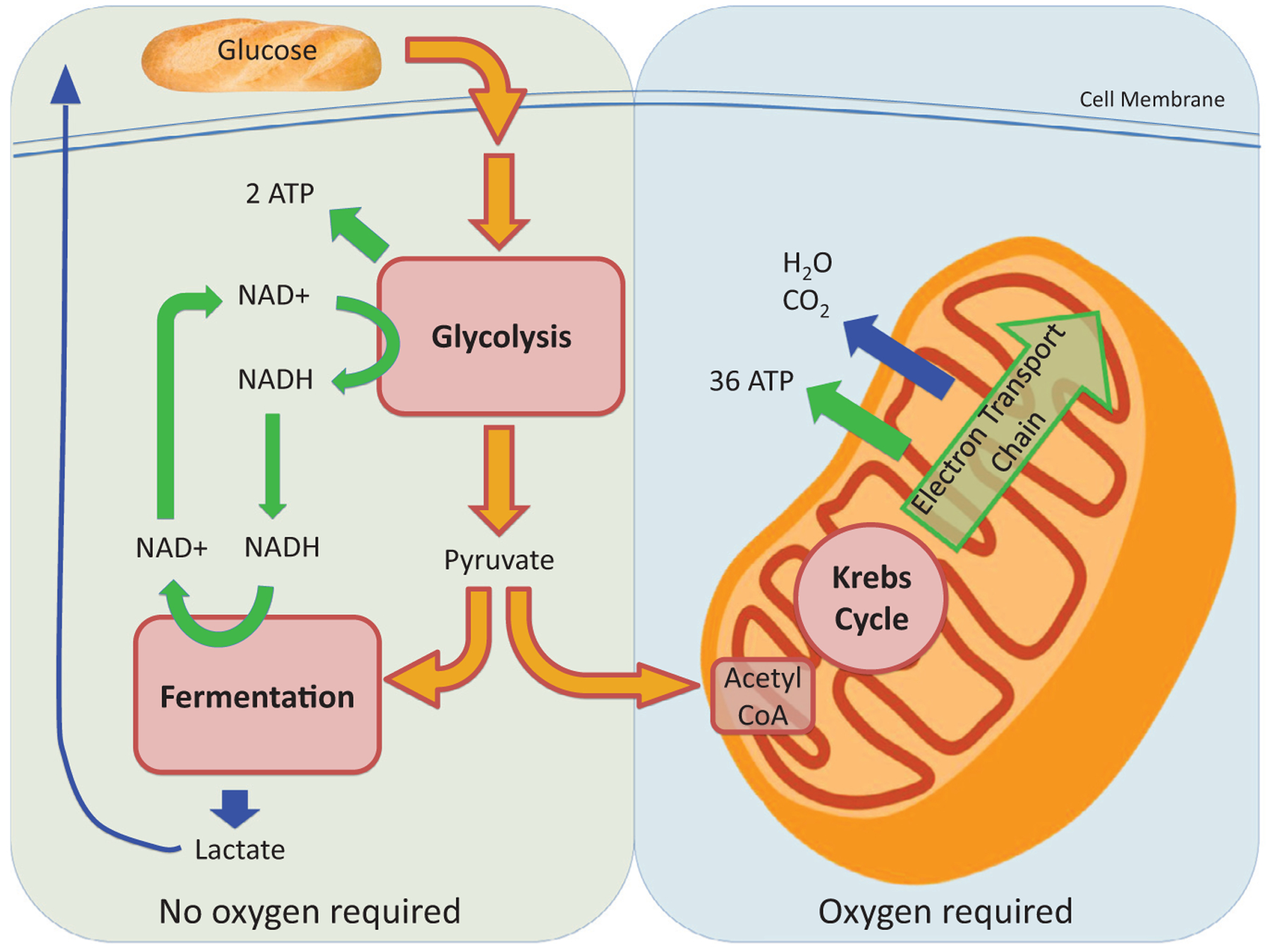
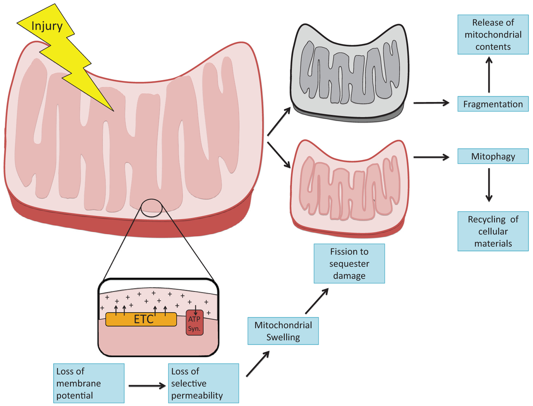
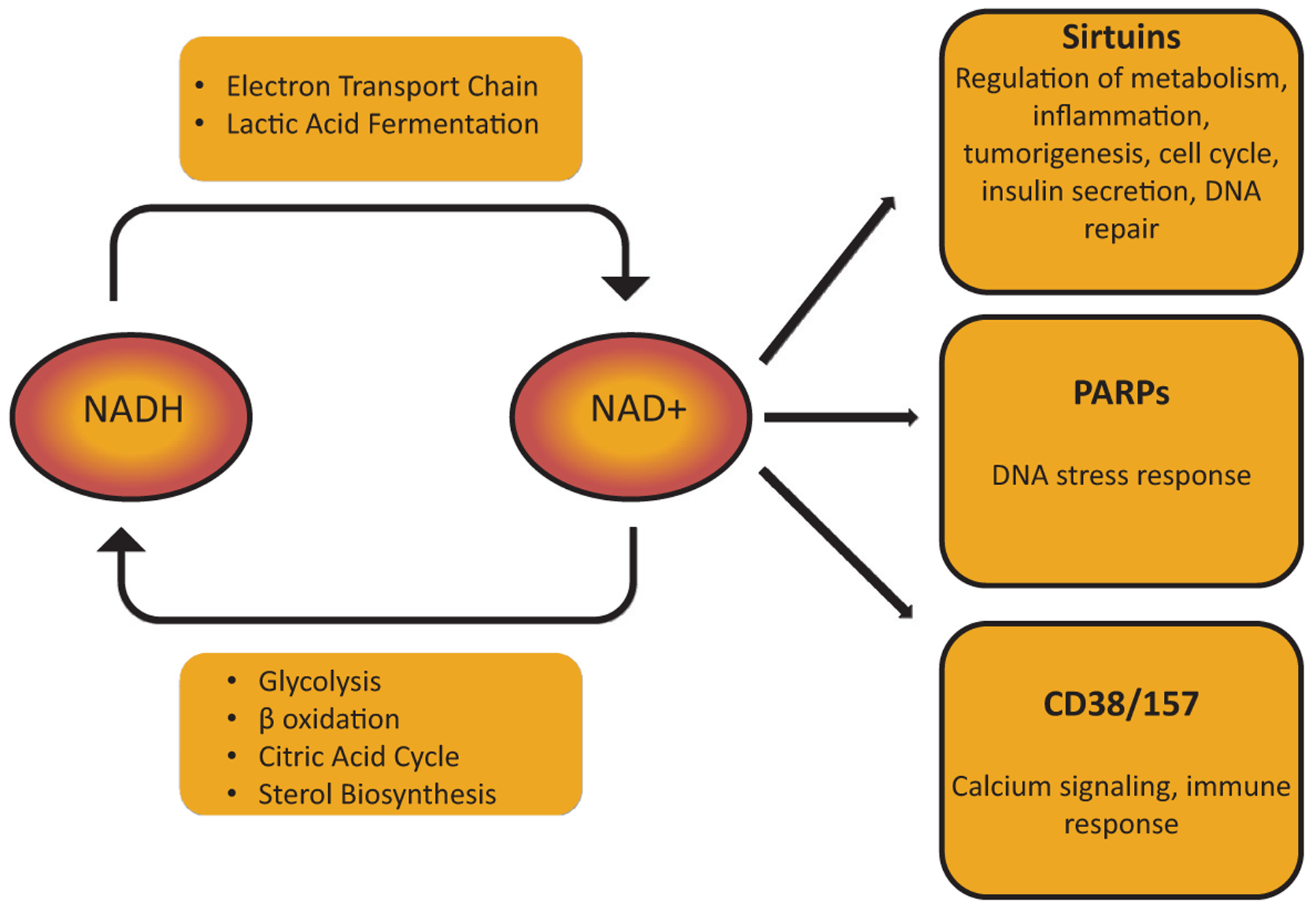
The kidney is a highly metabolic organ that requires substantial adenosine triphosphate for the active transport required to maintain water and solute reabsorption. Aberrations in energy availability and energy utilization can lead to cellular dysfunction and death. Mitochondria are essential for efficient energy production. The pathogenesis of acute kidney injury is complex and varies with different types of injury. However, multiple distinct acute kidney injury syndromes share a common dysregulation of energy metabolism. Pathways of energy metabolism and mitochondrial dysfunction are emerging as critical drivers of acute kidney injury and represent new potential targets for treatment. This review shows the basic metabolic pathways that all cells depend on for life; describes how the kidney optimizes those pathways to meet its anatomic, physiologic, and metabolic needs; summarizes the importance of metabolic and mitochondrial dysfunction in acute kidney injury; and analyzes the mitochondrial processes that become dysregulated in acute kidney injury including mitochondrial dynamics, mitophagy, mitochondrial biogenesis, and changes in mitochondrial energy metabolism.

Keywords: Acute kidney injury; metabolism; mitochondria.


Conflict of interest statement

Conflict of interest statement:

Samir M. Parikh is listed as an inventor on patent filings from Beth Israel Deaconess Medical Center, holds equity in Raksana Therapeutics, and has received consulting fees from Astellas, Cytokinesis, Mission Therapeutics, and Aerpio, where he serves on the Scientific Advisory Board.

Figures

Figure 1.
Figure 1.
Overview of cellular energy metabolism including glycolysis, fermentation, Krebs cycle, and ETC. These combined reactions produce ATP, which is critical for cellular function. Glycolysis and fermentation take place in the cytoplasm and do not require oxygen. Oxidative phosphorylation via the electron transport chain takes place in mitochondria and requires oxygen. Abbreviation: NADH, nicotinamide adenine dinucleotide.
Figure 2.
Figure 2.
Mitochondrial structural changes after injury. Different noxious stimuli, including inflammatory cytokines, ischemia-reperfusion, and toxins, injure mitochondria. Injury disrupts the normal vectorial pumping of protons across the inner mitochondrial membrane by enzymatic complexes in the ETC. Subsequent loss of membrane potential impairs selective permeability. As a result, mitochondria swell. Fission is induced to sequester the damage and safely dispose of injured mitochondria through mitophagy. Excessive fission, also known as mitochondrial fragmentation, arises during severe injury and is associated with the release of mitochondrial contents that potentiate inflammation and cell death. Abbreviation: ATP syn, ATP synthase.
Figure 3.
Figure 3.
Mitochondrial life cycle. The life cycle of mitochondria includes biogenesis of mitochondrial structural proteins from nuclear DNA and dynamic remodeling of the mitochondrial network via fission and fusion to maintain an optimally functioning mass of mitochondria within the cell. Mitophagy enables intracellular disposal of mitochondria and recycling of their contents for cellular needs. Excessive fission, typically in the setting of injury, leads to fragmentation and release of cytotoxic mitochondrial contents including pro-apoptotic factors, ROS, and inflammatory mtDNA. PGC1α is a key regulator of mitochondrial biogenesis. Fusion proteins mitofusin 1 (Mfn1) and mitofusin 2 (Mfn 2) are necessary for outer-membrane fusion, while optic atrophy 1 (Opa1) is necessary for inner-membrane fusion. Dynamin-related protein 1 (Drp1) is critical for mitochondrial fission. Bcl-2/adenovirus E1B 19 KDa-interacting protein (BNIP)3, FUN14 containing 1 (FUNDC1), and PTEN-induced kinase-1 (PINK-1) with parkin are all proteins that can trigger mitophagy.
Figure 4.
Figure 4.
Overview of NAD+ metabolism. In highly metabolically active cells such as the renal tubular epithelium, NAD+ regulates broad aspects of cellular metabolism. Reduction of NAD+ to NADH is required for glycolysis, fatty acid oxidation, the tricarboxylic acid (Krebs) cycle, and sterol biosynthesis. Oxidation of NADH back to NAD+ provides high-energy electrons in the ETC and generates lactic acid from pyruvate. NAD+ also can be used by enzymes that cleave NAD+ to generate Nam. These include PARPs, sirtuins, and cyclic ADP-ribose (cADPR) synthetases (CD38/CD157), each of which play crucial roles in cellular function.

References

    1. Sawhney S, Fraser SD. Epidemiology of AKI: utilizing large databases to determine the burden of AKI. Adv Chronic Kidney Dis. 2017;24:194–204. - PMC - PubMed
    1. Pavkov ME, Harding JL, Burrows NR. Trends in hospitalizations for acute kidney injury - United States, 2000–2014. MMWR Morb Mortal Wkly Rep. 2018;67:289–93. - PMC - PubMed
    1. Pagliarini DJ, Calvo SE, Chang B, et al. A mitochondrial protein compendium elucidates complex I disease biology. Cell. 2008; 134:112–23. - PMC - PubMed
    1. Harris SI, Balaban RS, Mandel LJ. Oxygen consumption and cellular ion transport: evidence for adenosine triphosphate to O2 ratio near 6 in intact cell. Science. 1980;208:1148–50. - PubMed
    1. Kurnik BR, Weisberg LS, Kurnik PB. Renal and systemic oxygen consumption in patients with normal and abnormal renal function. J Am Soc Nephrol. 1992;2:1617–26. - PubMed

Publication types