. 2020 Apr;8(1):e000676.
doi: 10.1136/jitc-2020-000676.

Inhibition of TGF- β- receptor signaling augments the antitumor function of ROR1-specific CAR T-cells against triple-negative breast cancer

Affiliations

Inhibition of TGF- β- receptor signaling augments the antitumor function of ROR1-specific CAR T-cells against triple-negative breast cancer

Tanja Stüber et al. J Immunother Cancer. 2020 Apr.

Erratum in

Abstract

Background: Immunotherapy with chimeric antigen receptor (CAR)-engineered T-cells is effective in some hematologic tumors. In solid tumors, however, sustained antitumor responses after CAR T-cell therapy remain to be demonstrated both in the pre-clinical and clinical setting. A perceived barrier to the efficacy of CAR T-cell therapy in solid tumors is the hostile tumor microenvironment where immunosuppressive soluble factors like transforming growth factor (TGF)-β are thought to inhibit the cellular immune response. Here, we analyzed whether CAR T-cells specific for the receptor tyrosine kinase-like orphan receptor 1 (ROR1) antigen, that is frequently expressed in triple-negative breast cancer (TNBC), are susceptible to inhibition by TGF-β and evaluated TGF-β-receptor signaling blockade as a way of neutralizing the inhibitory effect of this cytokine.

Methods: CD8+ and CD4+ ROR1-CAR T-cells were prepared from healthy donors and their antitumor function analyzed using the TNBC cell line MDA-MB-231 in vitro and in a microphysiologic 3D tumor model. Analyses were performed in co-culture assays of ROR1-CAR T-cells and MDA-MB-231 cells with addition of exogenous TGF-β.

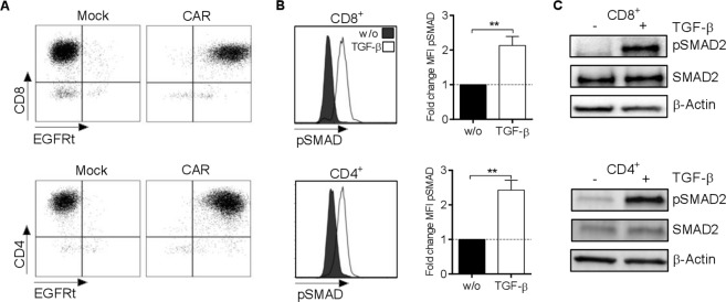
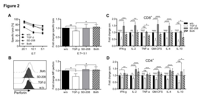
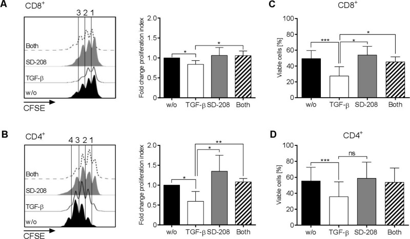
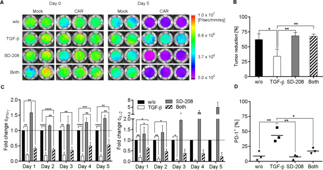
Results: The data show that exposure to TGF-β engages TGF-β-receptor signaling in CD8+ and CD4+ ROR1-CAR T-cells as evidenced by phosphorylation of small mothers against decapentaplegic homolog 2. In the presence of TGF-β, the cytolytic activity, cytokine production and proliferation of ROR1-CAR T-cells in co-culture with MDA-MB-231 TNBC cells were markedly impaired, and the viability of ROR1-CAR T-cells reduced. Blockade of TGF-β-receptor signaling with the specific kinase inhibitor SD-208 was able to protect CD8+ and CD4+ ROR1-CAR T-cells from the inhibitory effect of TGF-β, and sustained their antitumor function in vitro and in the microphysiologic 3D tumor model. Combination treatment with SD-208 also led to increased viability and lower expression of PD-1 on ROR1-CAR T-cells at the end of the antitumor response.

Conclusion: We demonstrate the TGF-β suppresses the antitumor function of ROR1-CAR T-cells against TNBC in preclinical models. Our study supports the continued preclinical development and the clinical evaluation of combination treatments that shield CAR T-cells from TGF-β, as exemplified by the TGF-β-receptor kinase inhibitor SD-208 in this study.

Keywords: breast neoplasms; immunotherapy; receptors, chimeric antigen.


Conflict of interest statement

Competing interests: None declared.

Figures

Figure 1
Figure 1
Transforming growth factor (TGF)-β induces small mothers against decapentaplegic homolog (SMAD) 2 phosphorylation in CD8+ and CD4+ receptor tyrosine kinase-like orphan receptor 1 (ROR1) chimeric antigen receptor (CAR) T-cells. (A) Phenotype of CD8+ (upper panel) and CD4+ (lower panel) CAR T-cells. Truncated epidermal growth factor receptor (EGFRt) is a detection marker encoded in cis with the CAR-transgene. (B, C) Analysis of SMAD2 phosphorylation in CD8+ (upper panel) and CD4+ (lower panel) ROR1-CAR T-cells assessed by intracellular flow cytometry staining (B) and Western blotting (C) under untreated control conditions and after incubation with TGF-β (10 ng/mL). (B) The histograms show data from a representative experiment. The right diagram shows fold change in SMAD2 phosphorylation as compared with untreated cells. Data shown are mean values±SD from n=3 independent experiments with **p<0.01 by Student’s t-test.
Figure 2
Figure 2
Effects of transforming growth factor (TGF)-β and SD-208 on cytolytic activity and cytokine production of receptor tyrosine kinase-like orphan receptor 1 (ROR1)-chimeric antigen receptor (CAR) T-cells. (A–C) ROR1-CAR T-cells were pretreated either with SD-208 (1 µM) or/and TGF-β (10 ng/mL) or were left untreated. (A) Specific lysis of MDA-MB-231 by ROR1-CAR CD8+ T-cells at different E:T ratios after a 4-hour co-culture. The left diagram shows specific killing of one representative experiment. Lysis percentage values are normalized to that of the mock control T-cell. The right diagram shows fold change of cytotoxicity as compared with untreated cells from three independent experiments at E:T=5:1. (B) CD8+ ROR1-CAR T-cells were fixed, permeabilized, and stained for intracellular perforin. The histogram shows data from one representative experiment. The right diagram shows fold change of perforin mean fluorescence intensity (MFI) as compared with untreated cells (n=3). (C, D) Luminex multiplex bead assays were performed on supernatants obtained after a 24-hour co-culture with MDA-MB-231 at an E:T ratio of 4:1. Data shown are mean values±SD of fold change concentration for each cytokine compared with its counterpart cytokine secreted by untreated cells. Cytokine production of CD8+ (C) and CD4+ (D) T-cells were obtained from n=10 (except TNF-α n=4) and n=9 different donors, respectively. Statistical analysis was performed using Student’s t-test (A, B) or one-way analysis of variance (C, D). *p<0.05, **p<0.01, ***p<0.001, ****p<0.0001, ns=not significant.
Figure 3
Figure 3
Effects of transforming growth factor (TGF-β) and SD-208 on proliferation and viability of receptor tyrosine kinase-like orphan receptor 1 (ROR1)-chimeric antigen receptor (CAR) T-cells. (A–D) CD8+ and CD4+ ROR1-CAR T-cells were co-cultured for 72 hours with MDA-MB-231 cells at E:T=4:1 in the presence of 1 µM SD-208 or/and 10 ng/mL TGF-β or were left untreated. Proliferation of CAR T-cells was measured by carboxyfluorescein succinimidyl ester (CFSE)-dilution in CD8+ (A) and CD4+ (B) T-cells. The diagrams on the right show fold change of the proliferation index compared with untreated cells. Percentages of viable CD8+ (C) and CD4+ (D) T-cells were assessed by 7-Aminoactinomycin staining. Data shown are mean values±SD from three independent experiments with *p<0.05, **p<0.01, ***p<0.001 by Student’s t-test, ns=not significant.
Figure 4
Figure 4
Effects of transforming growth factor (TGF)-β and SD-208 on specific lysis and cytokine secretion by CD8+ chimeric antigen receptor (CAR) T-cells in a static 3D tumor model. Over 5-day assay period, 5×10⁴ mock or receptor tyrosine kinase-like orphan receptor 1 (ROR1)-CAR T-cells were allowed to invade in microphysiologic 3D MDA-MB-231 models under control conditions or in the presence of 10 ng/mL TGF-β or/and 1 µM SD-208. (A) Images depicting the bioluminescence signal from viable tumor cells on day 0 and day 5 for one representative T-cell donor. (B) Reduction in tumor burden based on the bioluminescence signal encompassing the entire surface of the cell crowns normalized to the signal of mock cells. (C) Fold change of interferon (IFN-γ) and interleukin (IL-2)concentrations in supernatants at different time points as compared with untreated cells. (D) Proportion of PD-1+ cells from living (7-Aminoactinomycin) and CD45+ cells collected from the supernatant on day 5. Data shown are mean values±SD from three independent experiments with *p<0.05, **p<0.01, ***p<0.001, ****p<0.0001 by one-way analysis of variance, ns=not significant.

References

    1. Schmid P, Adams S, Rugo HS, et al. . Atezolizumab and nab-paclitaxel in advanced triple-negative breast cancer. N Engl J Med 2018;379:2108–21. 10.1056/NEJMoa1809615 - DOI - PubMed
    1. Chien H-P, Ueng S-H, Chen S-C, et al. . Expression of Ror1 has prognostic significance in triple negative breast cancer. Virchows Arch 2016;468:589–95. 10.1007/s00428-016-1911-3 - DOI - PubMed
    1. Balakrishnan A, Goodpaster T, Randolph-Habecker J, et al. . Analysis of Ror1 protein expression in human cancer and normal tissues. Clin Cancer Res 2017;23:3061–71. 10.1158/1078-0432.CCR-16-2083 - DOI - PMC - PubMed
    1. Hudecek M, Lupo-Stanghellini M-T, Kosasih PL, et al. . Receptor affinity and extracellular domain modifications affect tumor recognition by ROR1-specific chimeric antigen receptor T cells. Clin Cancer Res 2013;19:3153–64. 10.1158/1078-0432.CCR-13-0330 - DOI - PMC - PubMed
    1. Baselga J, Rothenberg ML, Tabernero J, et al. . Tgf-Beta signalling-related markers in cancer patients with bone metastasis. Biomarkers 2008;13:217–36. 10.1080/13547500701676019 - DOI - PubMed

Publication types

MeSH terms

Substances