. 2020 Jul;50(8):1083-1094.
doi: 10.1007/s00247-020-04658-5. Epub 2020 Apr 17.

Structural and perfusion magnetic resonance imaging of congenital lung malformations

Affiliations

Structural and perfusion magnetic resonance imaging of congenital lung malformations

Christian J Kellenberger et al. Pediatr Radiol. 2020 Jul.

Abstract

Background: A radiation-free advanced imaging modality is desirable for investigating congenital thoracic malformations in young children.

Objective: To describe magnetic resonance imaging (MRI) findings of congenital bronchopulmonary foregut malformations and investigate the ability of lung MRI for their classification.

Materials and methods: This is a retrospective analysis of consecutive MRI examinations performed for suspected congenital lung anomalies in 39 children (median age: 3.8 months, range: 2 days-15 years). Morphological and perfusion findings were characterised on respiratory-gated fast spin echo and dynamic contrast-enhanced sequences obtained at 1.5 tesla. Abnormalities were classified independently by two readers and compared to an expert diagnosis based on pathology, surgery and/or other imaging.

Results: Main diagnoses included bronchopulmonary lesions in 33 patients, scimitar syndrome in 4 patients, pulmonary arteriovenous malformation and oesophageal duplication cyst in one patient each. Of 46 observed abnormalities, 44 (96%) were classified correctly with very good interobserver agreement (96% concordance rate). The 39 detected lung lesions included isolated overinflation (17/39, 44%), cystic pulmonary airway malformation (8/39, 21%), bronchopulmonary sequestration (7/39, 18%), bronchogenic cyst (4/39, 10%) and hybrid lesion (3/39, 8%). All lung lesions presented as perfusion defect at peak pulmonary enhancement. Non-cystic lesions showed a delayed peak (median delay: 2.8 s, interquartile range: 0.5 to 4.0 s) in relation to normal lung parenchyma.

Conclusion: A dedicated lung MRI protocol including respiratory compensated sequences, dynamic angiography and perfusion is able to reliably delineate parenchymal and vascular components of congenital bronchopulmonary foregut malformations. Therefore, MRI may be considered for comprehensive postnatal evaluation of congenital thoracic malformations.

Keywords: Bronchopulmonary malformation; Children; Lung; Magnetic resonance imaging; Perfusion imaging.


Conflict of interest statement

None

Figures
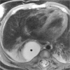
Fig. 1
Fig. 1
Oesophageal duplication cyst in a 3-month-old girl with foetal ultrasound diagnosis of cystic congenital pulmonary airway malformation. Axial fat-saturated T2-weighted image shows broad connection of the unilocular fluid-filled cyst (*) with the oesophageal wall
Fig. 2
Fig. 2
Bronchogenic cysts in an 8-week-old boy. a Coronal proton density MR image shows a single air-filled cyst (*) in the left upper lobe following thoraco-amniotic drainage in utero. b Axial T2-weighted image shows a second mediastinal fluid-filled cyst (**) with connection to the tracheal wall
Fig. 3
Fig. 3
Cystic congenital pulmonary airway malformation (CPAM) type 2 and isolated overinflation in an 8-month-old girl. a Axial T2-weighted image shows multiple air-filled cysts (arrow) in the anterior upper lobe segment and overinflation of the posterior upper lobe segment (arrowheads). b, c Axial image at peak enhancement (b) and parametric map (enhancement integral) (c) show the CPAM (2 in b, c) and isolated overinflation (3 in b, c) as perfusion defects. d Signal intensity – time curves show minimal perfusion of the CPAM (2 in d) and delayed enhancement of the overinflation (3 in d) in comparison to normal lung parenchyma (1 in b, c and d). ROI region of interest
Fig. 4
Fig. 4
Isolated overinflation (bronchial atresia) in an 11-month-old boy. a Axial T2-weighted image shows a hypointense lung area (arrowheads) with rarefication of the pulmonary markings, central fluid-filled round and linear bronchial structures (*, mucoceles), and some consolidation (arrow). b Coronal contrast-enhanced image at pulmonary peak enhancement shows the pulmonary lesion as a perfusion defect (arrowheads). c Coronal parametric map (enhancement integral). d Signal intensity – time curves. There is decreased perfusion of the overinflated area with pulmonary arterial peak (2 in c and d), while the consolidation (3 in c and d) shows delayed peak enhancement when compared to normal lung parenchyma (1 in c and d). ROI region of interest
Fig. 5
Fig. 5
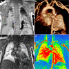
Bronchopulmonary sequestration in a 3-month-old boy. a Coronal proton density MR image shows consolidation in the left lower lobe (**) and prominent vessels with flow voids. b Left anterior oblique volume rendered angiographic view shows the lung lesion (**), its systemic arterial supply (arrowhead) from the descending aorta and venous drainage (arrow) to the left lower pulmonary vein. c, d Coronal image at peak pulmonary enhancement (c) shows a perfusion defect (**) and parametric map (enhancement integral) (d) shows increased perfusion (**) compared to normal lung (* in c and d)

References

    1. Langston C. New concepts in the pathology of congenital lung malformations. Semin Pediatr Surg. 2003;12:17–37. doi: 10.1016/S1055-8586(03)70004-3. - DOI - PubMed
    1. Newman B. Congenital bronchopulmonary foregut malformations: concepts and controversies. Pediatr Radiol. 2006;36:773–791. doi: 10.1007/s00247-006-0115-4. - DOI - PubMed
    1. Burge D, Wheeler R. Increasing incidence of detection of congenital lung lesions. Pediatr Pulmonol. 2010;45:103. doi: 10.1002/ppul.21150. - DOI - PubMed
    1. Bush A. Congenital lung disease: a plea for clear thinking and clear nomenclature. Pediatr Pulmonol. 2001;32:328–337. doi: 10.1002/ppul.1126. - DOI - PubMed
    1. Barth RA. Imaging of fetal chest masses. Pediatr Radiol. 2012;42(Suppl 1):S62–S73. doi: 10.1007/s00247-011-2171-7. - DOI - PubMed