

Case Reports
doi: 10.1016/j.jvir.2019.11.019.
Epub 2020 Apr 15.
Arteriovenous Graft Delamination and Dissection as a Cause of Graft Dysfunction
Affiliations
- PMID: 32305246
- PMCID: PMC7586731
- DOI: 10.1016/j.jvir.2019.11.019
Item in Clipboard
Case Reports
Arteriovenous Graft Delamination and Dissection as a Cause of Graft Dysfunction
J Vasc Interv Radiol.
2020 May.
No abstract available
Conflict of interest statement
Dr. Kim has received personal fees from Applied Clinical Intelligence (Bala Cynwyd, Pennsylvania) and Humacyte (Durham, North Carolina). None of the other authors have identified a conflict of interest.
Figures
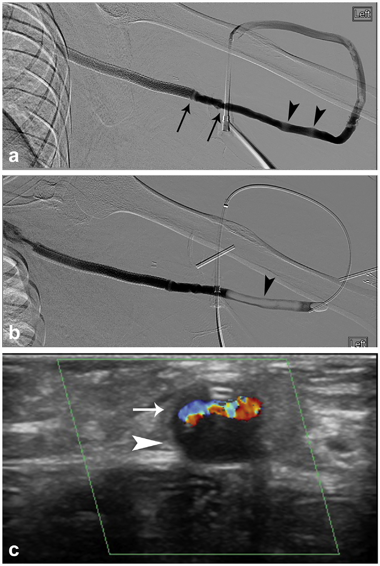
A 25-year-old female presented with thrombosis of a left arm Hybrid AV graft (W.L. Gore). (a) Following thrombectomy, graft venography demonstrated 2 mild focal stenoses near the graft-stent graft interface (arrows) and 2 apparently mild filling defects in the venous limb of the graft (arrowheads). (b) After occlusion was performed, using balloon pull thrombectomy with contrast injection with the balloon inflated, the filling defects markedly changed in appearance (arrowhead). After the occlusion balloon was removed, the apparent filling defects changed in appearance yet again (not shown). (c) Transverse color Doppler ultrasonography imaging of the graft demonstrates that the patent true lumen (arrow) is compressed by the false lumen that contains stagnant or thrombosed blood (arrowhead).
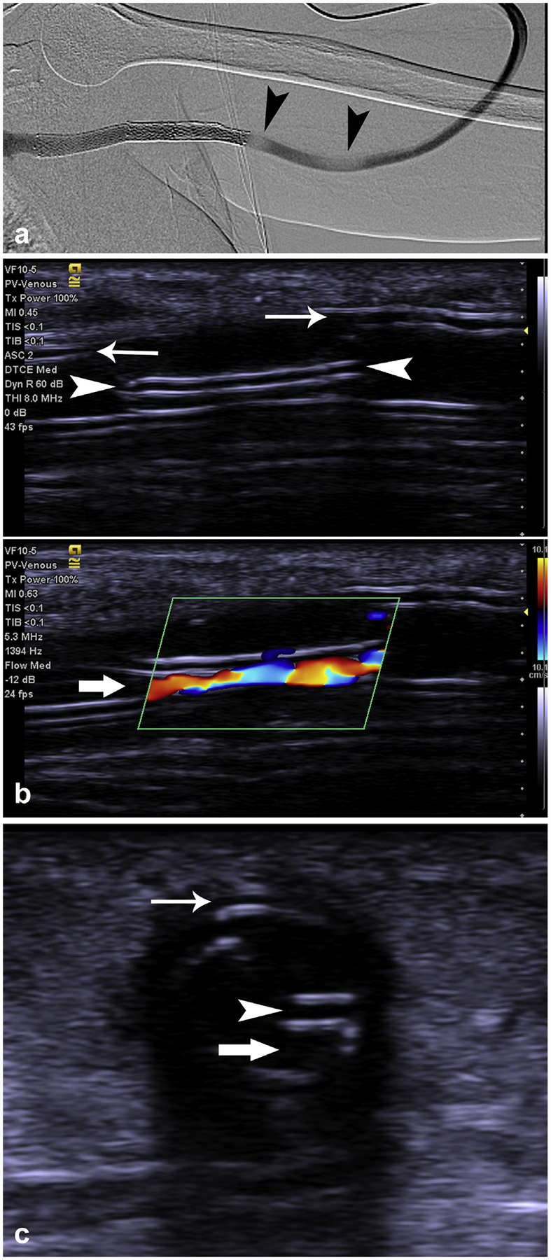
A 30-year-old female presented with elevated AVG pressure within her AV graft (Accuseal, W.L. Gore). (a) Angiography demonstrated apparent filling defects within the venous limb of the graft (arrowheads). (b) Longitudinal grayscale and corresponding color Doppler ultrasonography demonstrated disruption and displacement of the inner and middle layers of the graft (arrowheads) from the remainder of the graft (arrows), resulting in at least 80% narrowing of the true lumen (block arrow). (c) Transverse grayscale ultrasonography at the same location, shows dissected layers (arrowhead), native graft (arrow), and the true lumen (block arrow). AVG = arteriovenous graft.
References
-
- Roy-Chaudhury P, Kelly BS, Miller MA, et al. Venous neointimal hyperplasia in polytetrafluoroethylene dialysis grafts. Kidney Int 2001; 59:2325–2334. - PubMed
Publication types
MeSH terms
Grants and funding
LinkOut - more resources
Full Text Sources
Medical
