Review
. 2020 Aug;1868(8):140428.
doi: 10.1016/j.bbapap.2020.140428. Epub 2020 Apr 17.

Cathepsin B in neurodegeneration of Alzheimer's disease, traumatic brain injury, and related brain disorders

Affiliations
Review

Cathepsin B in neurodegeneration of Alzheimer's disease, traumatic brain injury, and related brain disorders

Vivian Hook et al. Biochim Biophys Acta Proteins Proteom. 2020 Aug.

Abstract

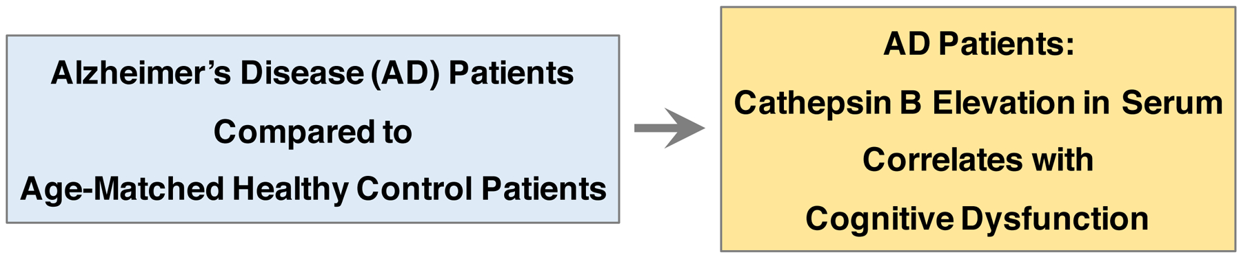
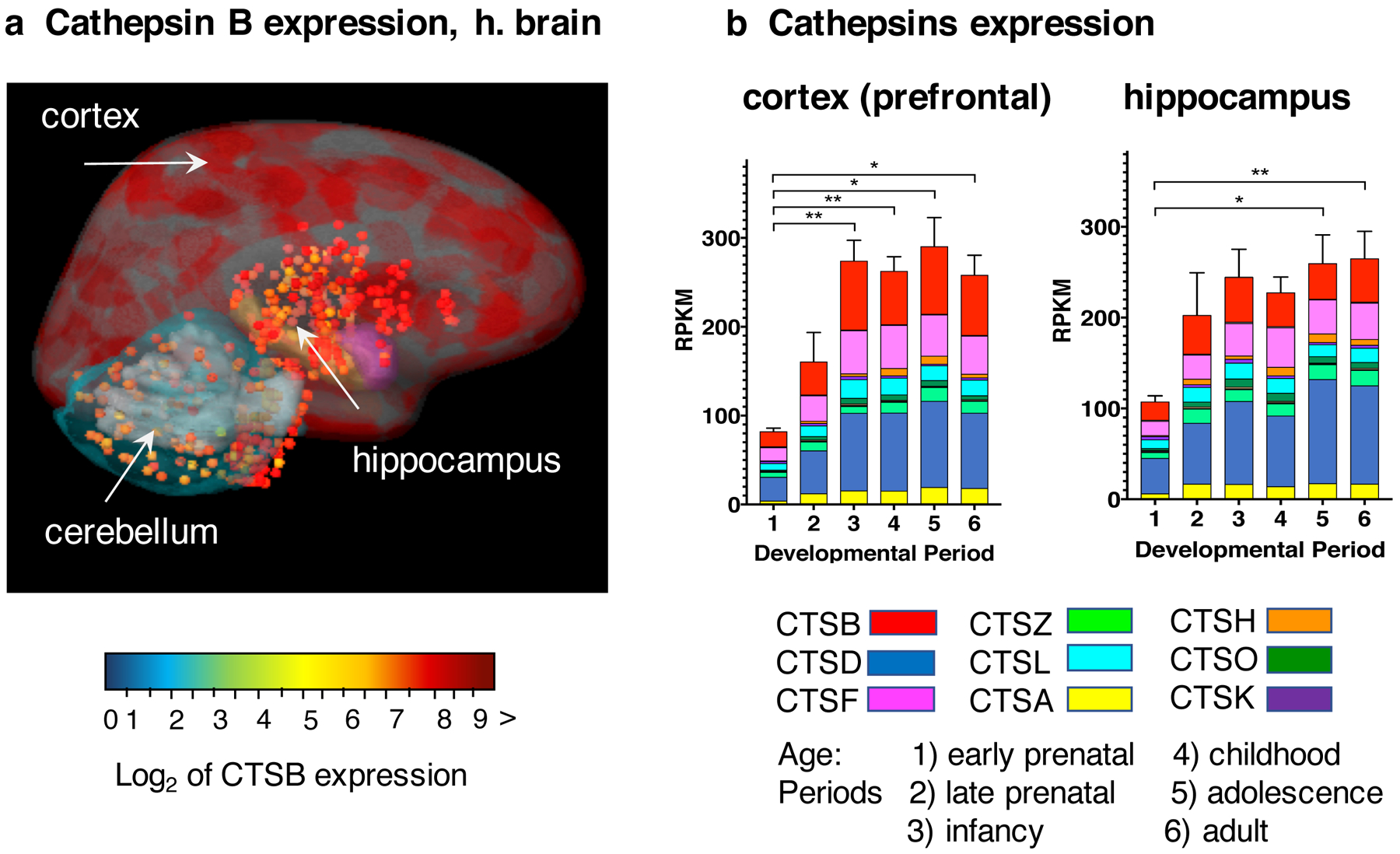
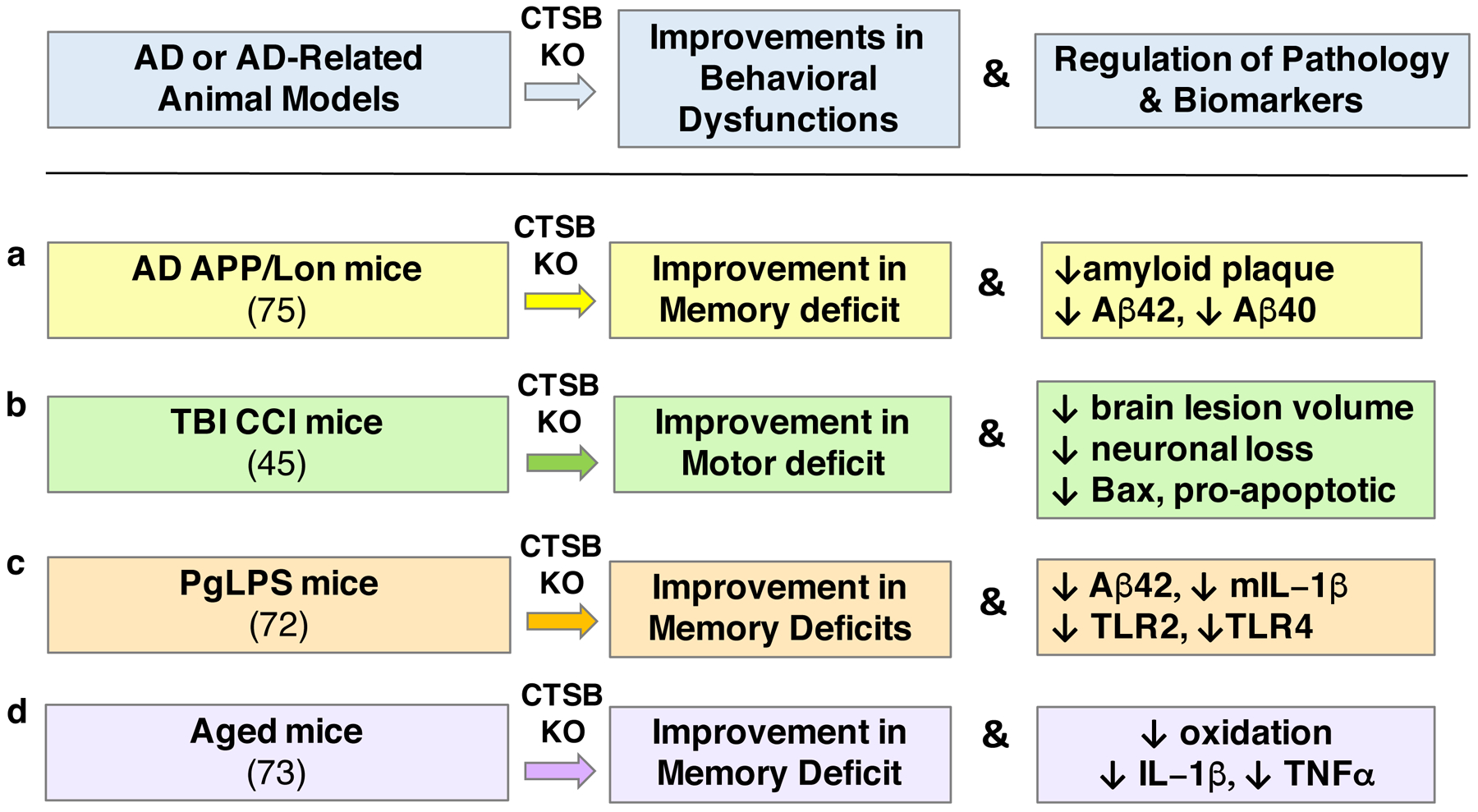
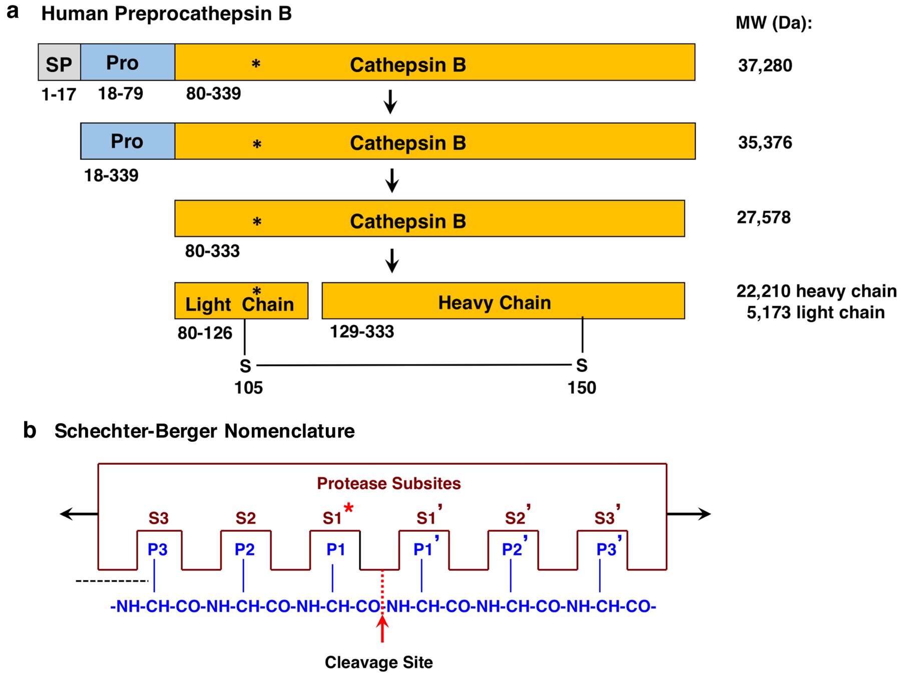
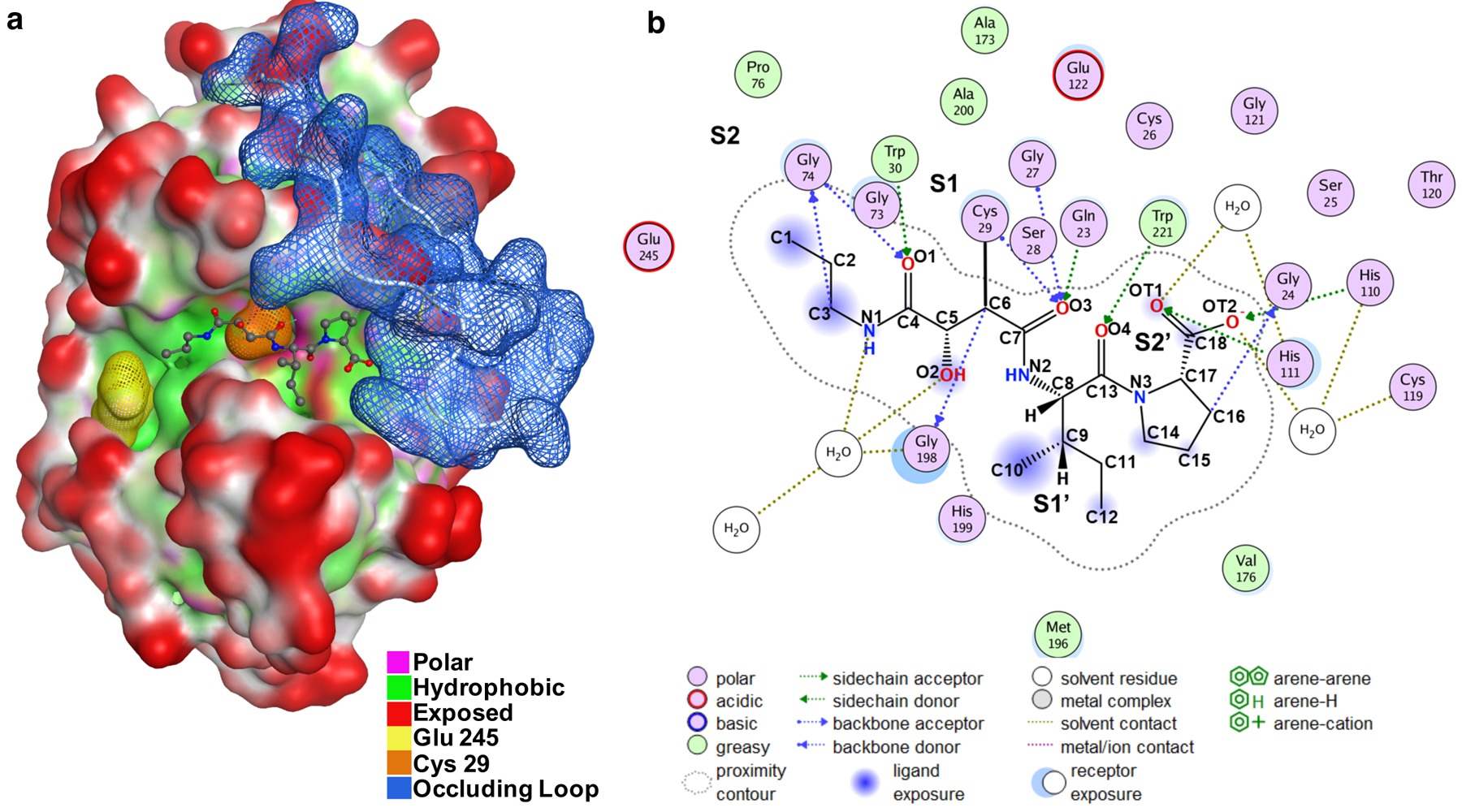
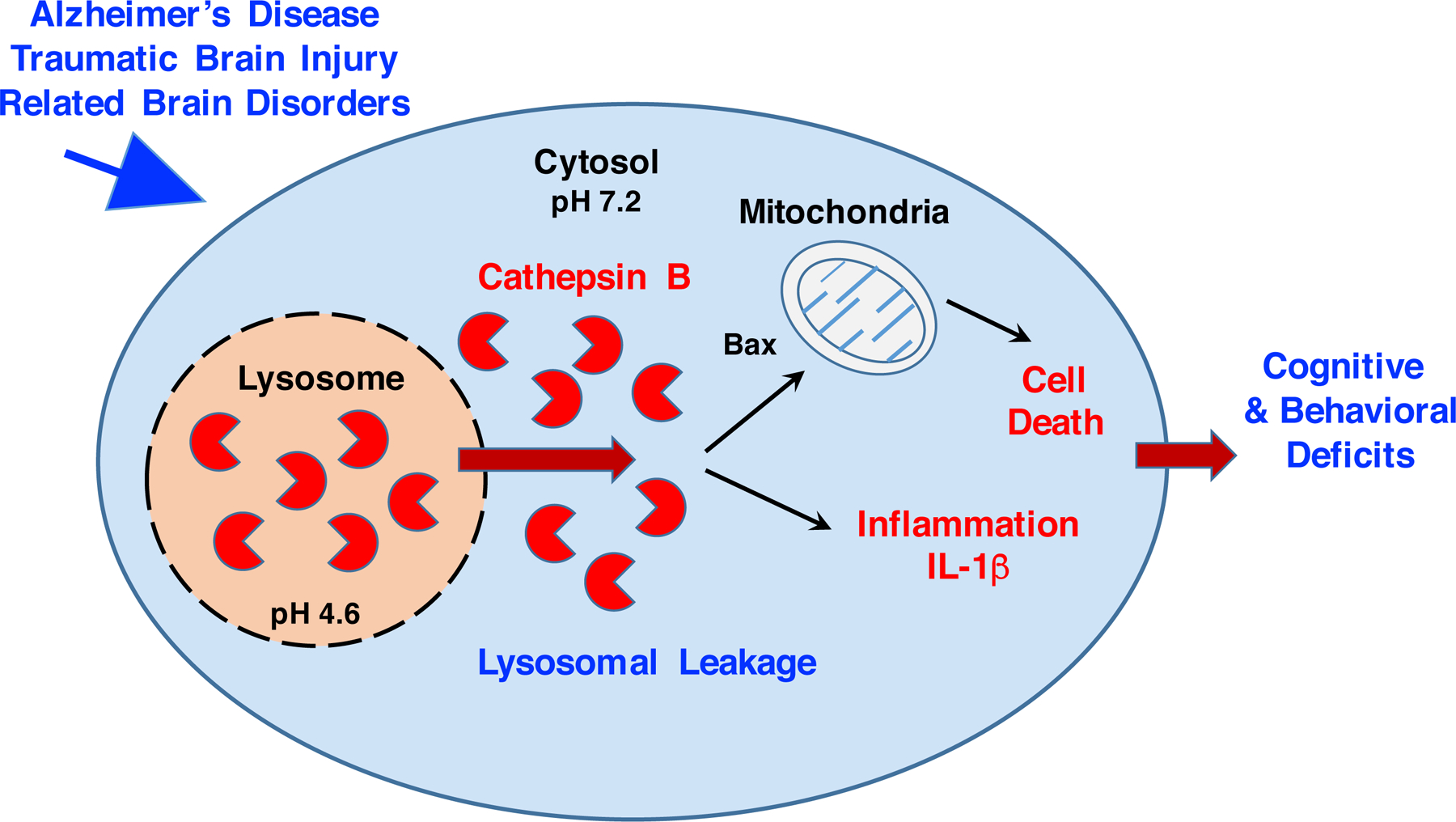
Investigations of Alzheimer's disease (AD), traumatic brain injury (TBI), and related brain disorders have provided extensive evidence for involvement of cathepsin B, a lysosomal cysteine protease, in mediating the behavioral deficits and neuropathology of these neurodegenerative diseases. This review integrates findings of cathepsin B regulation in clinical biomarker studies, animal model genetic and inhibitor evaluations, structural studies, and lysosomal cell biological mechanisms in AD, TBI, and related brain disorders. The results together indicate the role of cathepsin B in the behavioral deficits and neuropathology of these disorders. Lysosomal leakage occurs in AD and TBI, and related neurodegeneration, which leads to the hypothesis that cathepsin B is redistributed from the lysosome to the cytosol where it initiates cell death and inflammation processes associated with neurodegeneration. These results together implicate cathepsin B as a major contributor to these neuropathological changes and behavioral deficits. These findings support the investigation of cathepsin B as a potential drug target for therapeutic discovery and treatment of AD, TBI, and TBI-related brain disorders.

Keywords: Active site binding; Alzheimer's disease (AD); Behaviors; Biomarker; Brain; Cathepsin B; Cognition; Gene knockout; Human brain; Inhibitors; Lysosomal leakage; Memory; Neurodegeneration; Pathology; Traumatic brain injury (TBI).


Conflict of interest statement

Declaration of Competing Interest None

Figures

Figure 1.
Figure 1.. Elevation of cathepsin B in serum correlates with cognitive dysfunction in Alzheimer’s disease patients.
Cathepsin B levels were significantly elevated by ~50% in serum from AD patients compared to age-matched control patients, shown by studies of Sun et al., 2015 (29). Furthermore, the increased cathepsin B in serum was significantly correlated with cognitive dysfunction, measured the Mini-Mental State Exam which tests for cognitive functions of memory, attention, language, and orientation (29).
Figure 2.
Figure 2.. Expression of cathepsin B and lysosomal cathepsins in young and adult human brains.
a. Cathepsin B expression in human adult brain. Cathepsin B mRNA expression levels were determined by RNAseq analyses by the Allen Human Brain Atlas (https://human.brain-map.org/). The log2 normalized expression values of cathepsin B mRNA levels are illustrated overlaid on an MRI image of adult human brain, in color-coded ranges of high (dark red), medium (yellow), and lower (green-blue) levels of expression. The cortex regions display very high levels of cathepsin B expression and the hippocampus shows high levels of expression. b. Expression of cathepsin B and lysosomal cathepsin proteases in human brain regions at young through adult ages. Gene expression levels of cathepsin B and the majority of the lysosomal cathepsin proteases. Cathepsins D, F, Z, L, A, H, O, and K, representing the most abundantly expressed among 15 cathepsin proteases (40) are illustrated. Gene expression data are assessed as reads per kilobase million (RPKM) for six age periods of early prenatal, late prenatal, infancy, childhood, adolescence, and adult. Bar graphs show the total cathepsin gene expression as the average RPKM + s.e.m., with statistical significance (*p < 0.05, **p<0.01). Cathepsins B, F, and D are the most abundantly expressing cathepsins.
Figure 3.
Figure 3.. Cathepsin B gene knockout in animal models of AD, TBI, PgLPS, and aging.
Knockout (KO) of the cathepsin B gene (CTSB) results in alleviation of memory deficits in animal models of AD (75), PgLPS (72), and aging (73), and improvement in motor dysfunction in the CCI-TBI animal model (45), shown in panels a, b, c, and d, respectively. These improvements are accompanied by reduced neuropathology and reductions in biomarkers associated with the behavioral deficits. (a) In the APP/Lon mouse model of AD, CTSB KO results in substantial improvements in memory deficit measured by the Morris water maze test, and reduces amyloid plaque pathology with reduced brain levels of Aβ(–40) and Aβ(–42) (75), as well as reductions in pGlu-Aβ(–40) and pGlu-Aβ(–42) (77). (b) In the controlled cortical impact (CCI) model of traumatic brain injury (TBI), CTSB KO results in amelioration of motor deficits and allevation of brain tissue lesion pathology, combined with reduced neuronal loss and decreased pro-apoptotic Bax levels (45). (c) In mice suffering from cognitive decline due to infection by lipopolysaccharide from Porphyromonas gingivalis (PgLPS), CTSB KO results in improvement in memory deficit and reductions in Aβ42 and pro-inflammatory IL-1β, TLR2, and TLR4 (72). (d) CTSB KO in aged mice improves cognitive dysfunction, and ameliorates age-dependent increases in oxidation, IL-1β, and TNFα (73).
Figure 4.
Figure 4.. Processing of preprocathepsin B zymogen to active cathepsin B.
a. Preprocathepsin B processing to active protease. Preprocathepsin B (339 residues) (116) is translated from its mRNA at the rough endoplasmic reticulum (RER). The N-terminal signal sequence (SP, 17 residues) is removed at the RER by signal peptidase. The resultant procathepsin B (residues 18–333) is routed through the Golgi apparatus to lysosomes. The procathepsin B undergoes autoprocessing to remove the propeptide (Pro) to generate the active, mature cathepsin B (residues 80–333). Cathepsin B can undergo further processing into light and heavy chains linked by a disulfide bond. The active site Cys108 is indicated by the asterisk (*). Sequences of these forms of cathepsin B were obtained from the NCBI (196) and UniProt (197) protein sequence databases. It is noted that the NH2-terminal amino acid of mature cathepsin B (residue 80 of preprocathepsin B) may be referred as residue 1, with active site Cys29. b. Schechter-Berger Nomenclature for protease and peptide interactions at the substrate cleavage site. According to the Schechter-Berger nomenclature (123), protease and substrate interactions are defined by the enzyme (i) non-prime subsites of S1, S2, etc. which are located at the N-terminal side of the cleaved scissile bond, and the (ii) prime subsites of S1’, S2’, etc. which are located at the C-terminal side of the cleaved peptide bond. Amino acid residues located at the cleaved scissile bond (P1-↓P1’) are represented by P1, P2, and Pn residues located at the C-terminal side of the cleavage site, and by P1’, P2’, and Pn’ residues located at the N-terminal side of the scissile bond. The active site Cys108, shown by the asterisk (*), resides in the S1 subsite of the enzyme.
Figure 5.
Figure 5.. Interaction of the CA-074 inhibitor to the active site pocket of cathepsin B.
a. Structure of the cathepsin B/CA-074 complex. The structure of bovine cathepsin B complexed with CA-074 has been determined by x-ray crystallography by Yamamoto et al., 2000 (100); the PDB ID of this structural data of cathepsin B complexed with CA-074 is ID 1QdQ. This structural data was utilized by us with the Molecular Operating Environment (MOE) (130, 131) for visualization of the interacting features of CA-074 with the cathepsin B structure. The catalytic residue Cys29 (orange) is part of an extended substrate binding groove that is mostly hydrophobic (green) with some polar regions (pink). Using the Schechter and Berger nomenclature (123), Glu245 (yellow) sits in the S2 pocket and the occluding loop blue is orientated close to the S2ʹ pocket. The CA-074 inhibitor is orientated in the substrate binding groove and shown to extend from the S2 to S2ʹ pockets. b. Illustration of two-dimensional (2D) CA-074 inhibitor and cathepsin B interactions. Interactions of CA-074 with cathepsin B (bovine) were determined from their crystal structure (100); CA-074 interacts with S2’ to S2 subsites of the enzyme. The S2’ subsite interacts with the C-terminal Pro of CA-074 with His110 and His111 of the occluding loop by hydrogen bonding; the S2’ subsite involves Gln23, Gly24, His110, His111 of the occluding loop domain of cathepsin B. The occluding loop is located in manner to block substrate accessibility to Sn’ subsites (n > 3). The S1’ subsite interacts with the Ile of the Ile-Pro C-terminal region of CA-074; the S1’ site is composed of a hydrophobic core of Val176, Leu181, Met196, His199, and Trp221 which holds the Ile side chain of CA-074 through hydrogen bonding with Trp221. The S1 subsite contains the active site Cys29 which forms a covalent bond with the C6 oxirane carbon of CA-074; the S1 pocket is composed of Cys29, Gly27, Gly74, and Gly198. The S2 subsite consists of Glu245, Pro76, Ala173, and Ala200 which form a hydrophobic pocket which accommodates the propyl chain of CA-074. These features of CA-074 covalent linkage to enzyme and interactions with the substrate binding pocket in the vicinity of the occluding loop provide the basis for selective CA-074 inhibition of cathepsin B.
Figure 6.
Figure 6.. Lysosomal leakage results in cytosolic cathepsin B and activation of cell death neurodegeneration and inflammation in AD, TBI, and related.
Lysosomal leakage occurs in AD (–163), TBI (166, 167), and related brain disorders (–176). Lysosomal leakage results from membrane permeabilization, resulting in relocation of cathepsin B from the lysosome to the cytosol. Cathepsin B is active at the neutral pH 7.2 of the cytosol (–186), which differs from the acidic pH 4.6 within lysosomes (–183). Cathepsin B proteolysis of cytosolic substrates initiates apoptotic cell death (–146) and activation of IL-1β production in inflammation (–155).

References

    1. Montine TJ, Koroshetz WJ, Babcock D, Dickson DW, Galpern WR, Glymour MM, Greenberg SM, Hutton ML, Knopman DS, Kuzmichev AN, Manly JJ, Marder KS, Miller BL, Phelps CH, Seeley WW, Sieber BA, Silverberg NB, Sutherland M, Torborg CL, Waddy SP, Zlokovic BV, Corriveau RA; ADRD 2013 Conference Organizing Committee. Recommendations of the Alzheimer’s disease-related dementias conference. Neurology. 2014. August 26;83(9):851–60. - PMC - PubMed
    1. Corriveau RA, Koroshetz WJ, Gladman JT, Jeon S, Babcock D, Bennett DA, Carmichael ST, Dickinson SL, Dickson DW, Emr M, Fillit H, Greenberg SM, Hutton ML, Knopman DS, Manly JJ, Marder KS, Moy CS, Phelps CH, Scott PA, Seeley WW, Sieber BA, Silverberg NB, Sutherland ML, Taylor A, Torborg CL, Waddy SP, Gubitz AK, Holtzman DM. Alzheimer’s Disease-Related Dementias Summit 2016: National research priorities. Neurology. 2017. December 5;89(23):2381–2391. - PMC - PubMed
    1. Aisen PS, Cummings J, Jack CR Jr, Morris JC, Sperling R, Frölich L, Jones RW, Dowsett SA, Matthews BR, Raskin J, Scheltens P, Dubois B. On the path to 2025: understanding the Alzheimer’s disease continuum. Alzheimers Res Ther. 2017. August 9;9(1):60. - PMC - PubMed
    1. Ramalho J, Castillo M. Dementia resulting from traumatic brain injury. Dement Neuropsychol. 2015. Oct-Dec;9(4):356–368. - PMC - PubMed
    1. Brayne C. Traumatic brain injury and dementia. Lancet Psychiatry. 2018. May;5(5):383–384. - PubMed

Publication types

MeSH terms