. 2020 Apr:54:102738.
doi: 10.1016/j.ebiom.2020.102738.

Zika virus envelope nanoparticle antibodies protect mice without risk of disease enhancement

Affiliations

Zika virus envelope nanoparticle antibodies protect mice without risk of disease enhancement

Rahul Shukla et al. EBioMedicine. 2020 Apr.

Abstract

Background: Zika virus (ZIKV), an arbovirus capable of causing neurological abnormalities, is a recognised human pathogen, for which a vaccine is required. As ZIKV antibodies can mediate antibody-dependent enhancement (ADE) of dengue virus (DENV) infection, a ZIKV vaccine must not only protect against ZIKV but must also not sensitise vaccinees to severe dengue.

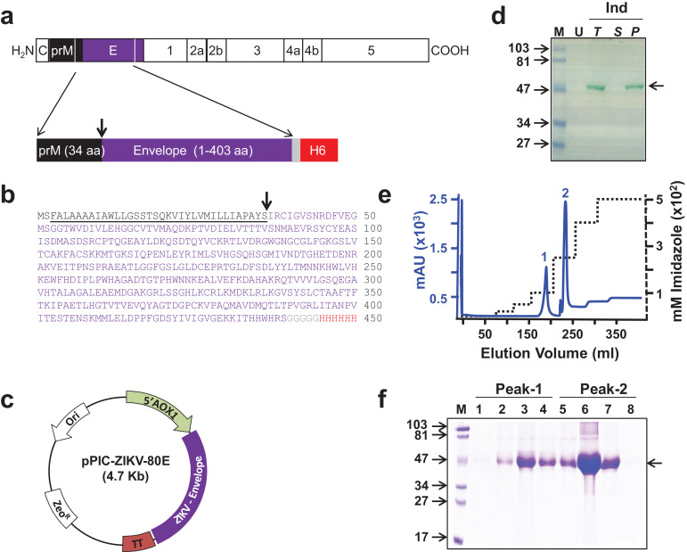
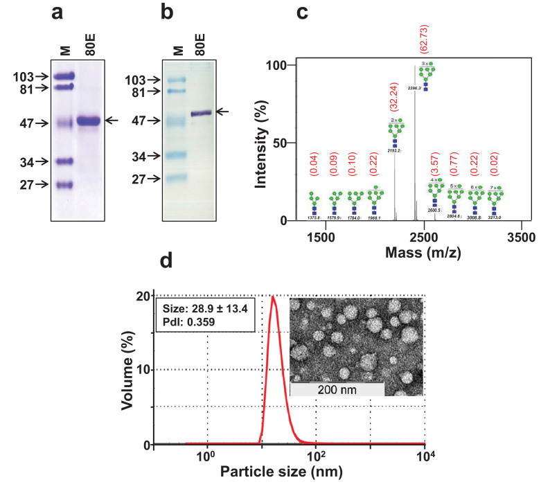
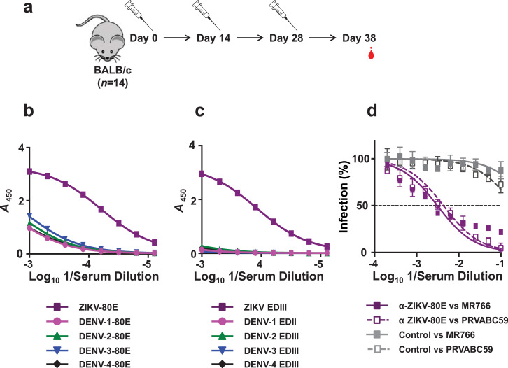
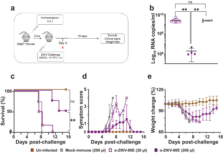
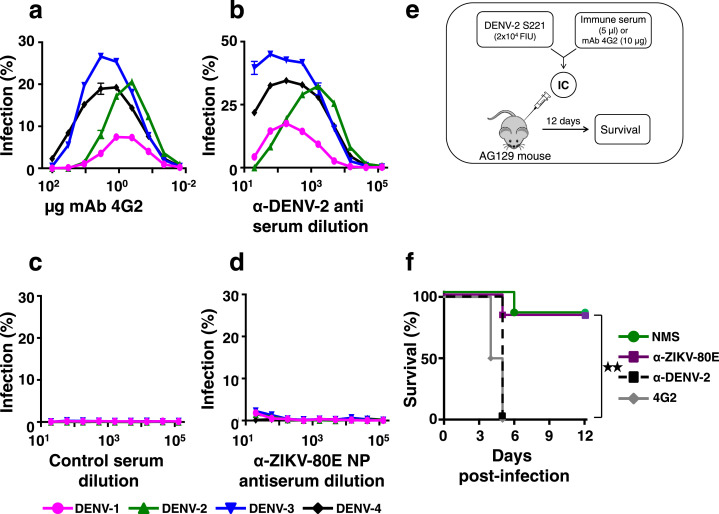
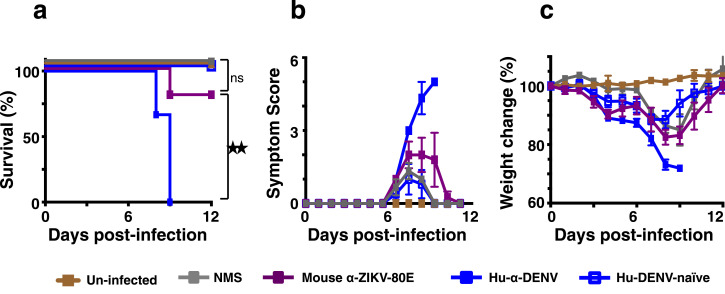
Methods: The N-terminal 80% of ZIKV envelope protein (80E) was expressed in Pichia pastoris and its capacity to self-assemble into particulate structures evaluated using dynamic light scattering and electron microscopy. Antigenic integrity of the 80E protein was evaluated using ZIKV-specific monoclonal antibodies. Its immunogenicity and protective efficacy were assessed in BALB/c and C57BL/6 Stat2-/- mice, respectively. Its capacity to enhance DENV and ZIKV infection was assessed in AG129 and C57BL/6 Stat2-/- mice, respectively.

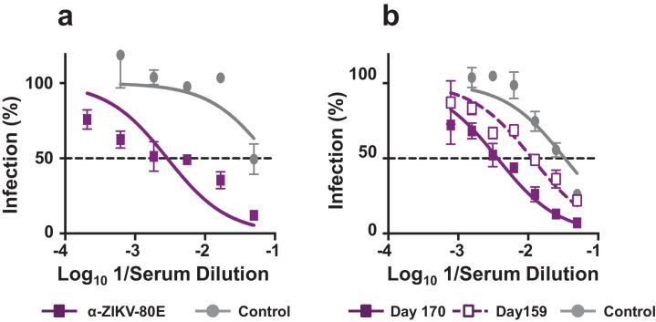
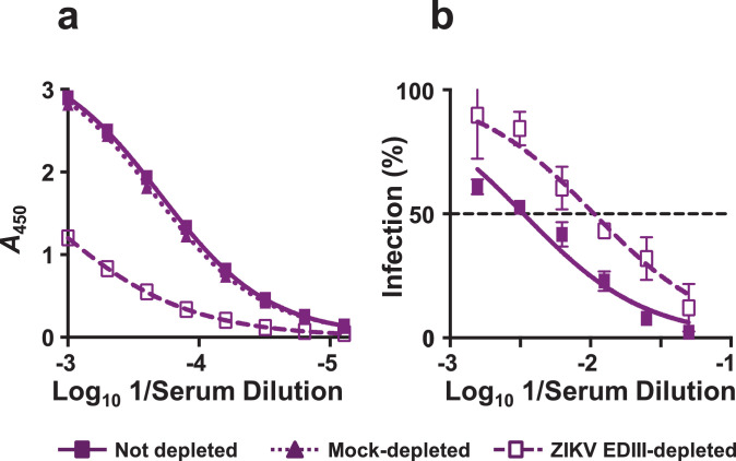
Findings: ZIKV-80E protein self-assembled into discrete nanoparticles (NPs), which preserved the antigenic integrity of neutralising epitopes on E domain III (EDIII) and elicited potent ZIKV-neutralising antibodies predominantly against this domain in BALB/c mice. These antibodies conferred statistically significant protection in vivo (p = 0.01, Mantel-Cox test), and did not exacerbate sub-lethal DENV-2 or ZIKV challenges in vivo.

Interpretation: Yeast-expressed ZIKV-80E, which forms highly immunogenic EDIII-displaying NPs, elicits ZIKV EDIII-specific antibodies capable of offering significant protection in vivo, without the potential risk of ADE upon subsequent DENV-2 or ZIKV infection. This offers a promising vaccine candidate for further development.

Funding: This study was supported partly by ICGEB, India, and by NIAID, USA.

Keywords: AG129; Antibody-dependent enhancement; C57BL/6 Stat2(−/−); Nanoparticles; Pichia pastoris;Dengue virus; VLPs; Zika virus vaccine.


Conflict of interest statement

Declaration of Competing Interest Nothing to disclose: GB, JAA; Grant from University Grants Commission, RKS; Grant from ICGEB: RS, VR, SS, NK; Grant from NIAID: JKL, FK; Patent 201911014359 pending: RS, RKS, VR, UA, SS, NK.

Figures

Fig. 1
Fig. 1
Design, expression, and purification of ZIKV-80E antigen. (a) Schematic representation of the ZIKV polyprotein. Proteins prM and E are indicated by black and purple boxes, respectively. The region of the polyprotein included in the antigen design is bounded by the two white lines in the C-terminal regions of prM and E. Shown below is the schematic representation of the design of the recombinant ZIKV-80E antigen consisting of the last 34 aa residues of prM (black box) and the first 403 aa residues of E (purple box). The C-terminally located grey and red boxes denote the pentaglycyl peptide linker and the hexa-histidine (H6) tag, respectively. (b) Predicted aa sequence of the recombinant ZIKV-80E antigen. The colour scheme corresponds to that shown in ‘a’. prM aa residues are underlined. The N-terminal dipeptide ‘MS’ was introduced during cloning. The downward arrow in panels ‘a’ and ‘b’ denotes the signal peptide cleavage site. (c) Map of the ZIKV-80E expression plasmid, pPIC-ZIKV-80E. The ZIKV-80E gene (ZIKV-Envelope) is inserted between the AOX1 promoter (5′ AOX1) on the 5′ side and the transcriptional terminator (TT) on the 3′ side. The vector carries the zeocin selection marker (ZeoR), which is functional in both E. coli and P. pastoris, and the E. coli origin of replication (Ori), for bacterial propagation. (d) Analysis of localisation of recombinant ZIKV-80E protein in P. pastoris. An aliquot of methanol-induced (Ind) culture of P. pastoris was lysed with glass beads and separated into supernatant (S) and pellet (P) fractions. Total (T) extract prepared from an equivalent aliquot of the induced culture, S, and urea-solubilised P fractions were run on SDS-polyacrylamide gel and subjected to Western blot analysis using mAb 24A12. T extract prepared from an equivalent aliquot of the un-induced (U) culture was analysed in parallel. Pre-stained protein markers were analysed in lane ‘M’. Their sizes (in KDa) are indicated to the left. The arrow on the right indicates the position of the recombinant ZIKV-80E protein. (e) Chromatographic profile of recombinant ZIKV-80E purification by Ni2+ affinity chromatography, starting from P fraction of induced cell lysate under denaturing conditions. The solid blue and the dotted black curves represent the profiles of UV absorbance (at 280 nm) and the imidazole step-gradient, respectively. Bound protein was eluted as two peaks (1 and 2). (f) Coomassie-stained SDS-polyacrylamide gel analysis of fractions corresponding to peaks 1 (lanes 1–4) and 2 (lanes 5–8) shown in panel ‘e’. Protein markers were analysed in lane ‘M’. Their sizes (in KDa) are indicated to the left. The arrow on the right indicates the position of the recombinant ZIKV E protein.
Fig. 2
Fig. 2
Physical characterisation of purified ZIKV-80E protein. (a) Coomassie-stained SDS-polyacrylamide gel analysis of pooled peaks 1 and 2 (from Fig. 1f) after dialysis. (b) Western blot analysis of the pooled peaks using mAb 24A12. In panels ‘a’ and ‘b’, protein markers were run in lanes ‘M’ and the purified ZIKV-80E protein was run in lanes marked ‘80E’. Their sizes (in KDa) are shown to the left of each panel. The arrow on the right, of both these panels, indicates the position of the ZIKV-80E protein. (c) The mass spectrometric N-glycan profile of recombinant ZIKV-80E protein, obtained using Bruker UltraFlex II MALDI-TOF mass spectrometer. The numbers in black above/across the peaks indicate the masses of the N-glycan species. The proposed structures of the N-glycan moieties, with the blue squares representing N-acetyl glucosamine and the green circles representing mannose residues, are shown for each of the peaks. The numbers in red shown in parentheses above the peaks denote the relative abundance (%) of the N-glycan moieties of the purified ZIKV-80E protein. (d) Analysis of volume distribution profile of particles in purified ZIKV-80E preparation. The left inset shows DLS parameter values, obtained based on size distribution by intensity, and the right inset depicts the transmission EM image of ZIKV-80E NPs (the scale is shown on the left lower edge).
Fig. 3
Fig. 3
Determination of ZIKV-80E NP-induced antibody titres in mice. (a) Schematic representation of the immunisation schedule. Mice (n = 14) were given i.p. injections on days 0, 14, and 28, and bled 10 days later for antibody titration. A single dose contained 20 µg NPs formulated with 500 µg alum. (b) Indirect ELISA titration curves obtained using pooled immune sera collected on day 38 using P. pastoris-produced purified 80E NPs of ZIKV (purple), DENV-1 (magenta), DENV-2 (green), DENV-3 (blue) and DENV-4 (black), as coating antigens. (c) Similar indirect ELISA titration curves of the same immune sera pool as in panel ‘b’, but using E. coli-expressed, purified EDIII (as MBP fusion) proteins, corresponding to the same set of five viruses listed above, as the coating antigens. Data in panels ‘b’ and ‘c’ are plotted as ELISA reactivity, in terms of absorbance at 450 nm (A450), as a function of serum dilution. Each data point represents the average of duplicates. The ELISA titration curves shown depict one of two independent experiments. (d) Determination of ZIKV nAb titres in ZIKV-80E NP-immunised mice using FACS neutralisation assay. Serial dilutions of pooled heat-inactivated immune sera collected on day 38 were titrated against ZIKV MR766 (solid purple curve) and ZIKV PRVABC59 (dashed purple curve). In parallel, mock-immune serum was similarly titrated against the same two ZIKV strains (MR766: solid grey curve; PRVABC59: dashed grey curve). Data depict ZIKV infection (%) as a function of serum dilution. The horizontal dashed line denotes 50% ZIKV infection.
Fig. 4
Fig. 4
Determination of recall nAb response in immunised mice using the ZIKV luciferase reporter assay. (a) Determination of ZIKV nAb titre in the same pooled immune serum as in Fig. 3d, but determined using ZIKV RVPs. The data are represented as percent infection (in terms of Renilla luciferase activity of ZIKV RVPs) as a function of immune serum dilution, with luciferase activity of ZIKV RVP in absence of immune serum taken to represent 100% infection. Virus neutralisation curves obtained using pooled sera from mock-immunised and ZIKV-80E NP-immunised mice are shown using solid grey and solid purple curves, respectively. (b) A subset (n = 5) of mice was bled ∼4 months after the 3rd immunisation dose (on day 159) and given a booster dose of alum-formulated ZIKV-80E NPs (4th dose) the next day to assess recall response. Ten days later (day 170) the mice were bled again. Sera pools corresponding to day 159 (dashed purple curve) and 170 (solid purple curve) were assayed for nAb titres as described in panel ‘a’. Mock-immune serum analysed in parallel is shown by the solid grey curve. The dashed horizontal line in both panels denotes 50% infection based on ZIKV RVP luciferase activity.
Fig. 5
Fig. 5
Depletion of EDIII-specific antibodies from anti-ZIKV-80E NP antiserum is accompanied by reduction in ZIKV nAb titres. (a) Pooled serum (n = 5) collected on day 170 from BALB/c mice immunised with 4 doses of ZIKV-80E NPs (on days 0, 14, 28, and 160) was analysed for ZIKV EDIII-specific antibody titres by indirect ELISA using MBP-ZIKV EDIII as the coating antigen. The pooled serum was tested either before (solid curve) or after depletion on amylose resin-bound MBP protein (dotted curve) or MBP-ZIKV EDIII (dashed curve). Data are plotted as ELISA reactivity, in terms of absorbance at 450 nm (A450), as a function of serum dilution. Each data point represents the average of duplicates. The ELISA titration curves shown depict one of two independent experiments. (b) The pooled serum described in panel ‘a’ was assayed for ZIKV nAb titres either before (solid curve) or after (dashed curve) depletion on amylose resin-bound MBP-ZIKV EDIII using ZIKV RVP neutralisation assay. Note that in this experiment, MBP-depleted serum was omitted as its total IgG titres did not change significantly after MBP depletion, as shown in panel ‘a’ (dotted curve). The data are represented as percent infection (in terms of Renilla luciferase activity of ZIKV RVPs) as a function of immune serum dilution, with luciferase activity of ZIKV RVP in absence of immune serum taken to represent 100% infection. The dashed horizontal line denotes 50% inhibition of ZIKV reporter activity.
Fig. 6
Fig. 6
Evaluation of the capacity of ZIKV-80E NP-induced antibodies to protect C57BL/6 Stat2/− mice against lethal ZIKV MR766 challenge. (a) Schematic depiction of the efficacy test using C57BL/6 Stat2−/- mice. Mice were passively administered (i.p.) with 20 µl anti-ZIKV-80E NP antiserum [‘α-ZIKV-80E (20 µl)’ group, n = 6], 200 µl anti-ZIKV-80E NP antiserum [‘α-ZIKV-80E (200 µl)’ group, n = 4], or 200 µl mock-immune serum [‘Mock immune (200 µl)’ group, n = 4] and challenged i.d. with ZIKV MR766 (103 PFU/mouse). A fourth group of mice, which received neither passive serum transfer nor the challenge virus (‘Un-infected’ group) was included for comparison. Mice were bled on day 4 post-challenge for ZIKV RNA determination. All mice were monitored for the criteria listed for up to 15 days. (b) Blood plasma viral RNA levels (ZIKV genome copies/ml plasma) in ZIKV MR766-challenged mice, determined by q-RTPCR on day 4 post-challenge as described (Methods S1). The different groups are designated by the following symbols: empty purple squares, ‘α-ZIKV-80E (20 µl)’ group; solid purple squares, ‘α-ZIKV-80E (200 µl)’ group; and solid grey squares, ‘Mock immune (200 µl)’ group. Error bars represent mean ± standard deviation. The twin star symbol denotes significant difference between viremia in the ‘α-ZIKV-80E (200 µl)’ group, compared to either the ‘α-ZIKV-80E (20 µl)’ group (p = 0.0044) or the ‘Mock immune (200 µl)’ group (p = 0.0017), based on unpaired t-test with Welch's correction. (c) Kaplan–Meier survival curves of the different mice groups described in ‘a’. Survival of mice in the ‘α-ZIKV-80E (200 µl)’ group was significantly higher than that in the ‘α-ZIKV-80E (20 µl)’ group (p = 0.0074) and ‘Mock-immune (200 µl)’ group (p = 0.010), but comparable to that in the ‘Un-infected’ group (p = 0.0888), based on the Log-Rank (Mantel–Cox) test. (d) Clinical progression (using a slightly modified scoring system described earlier [24]) of the same mice groups in ‘c’. (e) Body weight loss profiles of the same groups of mice during the course of the experiment. In panels ‘c-e’ the different mouse groups are indicated by: dashed purple curve, ‘α-ZIKV-80E (20 µl)’ group; solid purple curve, ‘α-ZIKV-80E (200 µl)’ group; solid grey curve, ‘Mock immune (200 µl)’ group; and sold brown curve, ‘Un-infected’ group.
Fig. 7
Fig. 7
Evaluation of DENV infection-enhancement by anti-ZIKV-80E NP antiserum. (a) Each of the four DENV serotypes was separately pre-incubated with different amounts of mAb 4G2 (0.01–100 µg), and then allowed to infect K562 cells for 24 h. The percentage of DENV-infected cells was determined by flow cytometry using mAb 2H2-Alexa 488 conjugate. Data depict infection profiles of DENV-1 (magenta), DENV-2 (green), DENV-3 (blue) or DENV-4 (black), as functions of mAb 4G2 concentration. (b) Similar experiment as in panel ‘a’, except that DENV pre-incubations were performed with pooled, serially diluted (10−1–10−5) immune serum, obtained from BALB/c mice immunised with DENV-2 S16803 strain (α-DENV-2 antiserum). (c) In vitro ADE experiment as in panel ‘b’, except that the murine polyclonal antiserum was from mock-immunised BALB/c mice (control serum); (d) Similar experiment as in panels ‘b’ and ‘c’, but using pooled immune serum from ZIKV-80E NP-immunised BALB/c mice (α-ZIKV-80E NP antiserum). (e) Schematic representation of the in vivo DENV-2 ADE experiment in AG129 mice (abbreviations: IC: immune complex; FIU: FACS infectious unit). (f) Kaplan–Meier survival curves for groups (n = 6) of AG129 mice which were intravenously administered with a sub-lethal dose of DENV-2 S221 (pre-incubated with NMS, green curve) or with ICs generated in vitro by pre-incubating the sub-lethal dose of DENV-2 S221 with mAb 4G2 (4G2, grey curve), murine anti-DENV-2 antiserum (α-DENV-2, black dashed curve) or with serum from BALB/c mice immunised with ZIKV-80E NPs (α-ZIKV-80E, purple curve). Survival was observed for 12 days post-challenge. Survival of mice in the α-ZIKV-80E and NMS groups were comparable to each other (p = 0.5282) and significantly higher (p = 0.0095) than that in the 4G2 or α-DENV-2 groups, based on the Log-Rank (Mantel–Cox) test. Statistical differences are indicated as not significant (ns) or as significant (twin star symbol).
Fig. 8
Fig. 8
Evaluation of ZIKV infection-enhancement by anti-ZIKV-80E NP antiserum. C57BL/6 Stat2−/− mice were inoculated i.p. with 20 µl dengue-positive human plasma (Hu-α-DENV, solid blue squares), 20 µl dengue-negative human plasma (Hu-DENV-naïve, empty blue squares), 20 µl naïve mouse serum (NMS, solid grey squares) or 20 µl anti-ZIKV-80E NP antiserum (mouse α-ZIKV-80E, solid purple squares) and then challenged with live ZIKV (PRVABC59, 5 × 103 PFU/mouse, i.d.) two hours later. One group of mice which received neither immune serum nor ZIKV inoculation (Un-infected, solid brown squares) was included for comparison. Mice were monitored for survival (panel ‘a’), clinical symptoms (scored as described , panel ‘b’) and weight loss (panel ‘c’) for a period of 12 days following virus challenge. Differences between the weights of mice in the ‘Hu-α-DENV’, ‘Hu-DENV-naïve’ and ‘mouse α-ZIKV-80E’ groups, for days 3, 4, 5, and 6, analysed using a one way ANOVA (Tukey's test for multiple comparisons), were statistically not significant (p>0.05). Survival in the ‘mouse α-ZIKV-80E’ group was significantly higher than that in ‘Hu-α-DENV of mice’ group (p = 0.027) and comparable to ‘NMS’ group (p = 0.371) and ‘Un-infected’ group (p = 0.438), based on Mantel–Cox test. Statistical differences are indicated as not significant (ns) or as significant (twin star symbol).

References

    1. Pierson T.C., Diamond M.S. Flaviviruses. In: Knipe DM, Howley PM, editors. Fields virology. 6 ed. Wolters Kluwer and Lippincott Williams & Wilkins; Philadelphia: 2013. pp. 747–794.
    1. Pierson T.C., Diamond M.S. The emergence of Zika virus and its new clinical syndromes. Nature. 2018;560:573–581. - PubMed
    1. Gatherer D., Kohl A. Zika virus: a previously slow pandemic spreads rapidly through the Americas. J Gen Virol. 2016;97:269–273. - PubMed
    1. Lazear H.M., Diamond M.S. Zika virus: new clinical syndromes and its emergence in the Western hemisphere. J Virol. 2016;90:4864–4875. - PMC - PubMed
    1. Musso D., Roche C., Robin E., Nhan T., Teissier A., Cao-Lormeau V.M. Potential sexual transmission of Zika virus. Emerg Infect Dis. 2015;21:359–361. - PMC - PubMed

MeSH terms

Substances