

Evaluation of microvascular network with optical coherence tomography angiography (OCTA) in branch retinal vein occlusion (BRVO)
- PMID: 32306978
- PMCID: PMC7169004
- DOI: 10.1186/s12886-020-01405-0
Evaluation of microvascular network with optical coherence tomography angiography (OCTA) in branch retinal vein occlusion (BRVO)
Abstract
Background: To evaluate changes of microvascular network of macular and peripapillary regions and to provide a quantitative measurement of foveal avascular zone (FAZ) in unilateral BRVO patients.
Methods: Forty-seven unilateral BRVO patients and forty-seven normal controls were enrolled. A 3*3 mm scan centered on fovea followed by a 4.5*4.5 mm scan centered on optic nerve head (ONH) were obtained in BRVO eyes, fellow eyes and control eyes of each individual using OCTA (Optovue Inc., Fremont, CA, USA). Vessel density (VD) in superficial (SVC) and deep vascular complex (DVC) of macula and radial peripapillary capillary (RPC) were automatically calculated. Parameters of FAZ region including size, perimeter, acircularity index (AI) and foveal vessel density 300 (FD-300) were measured.
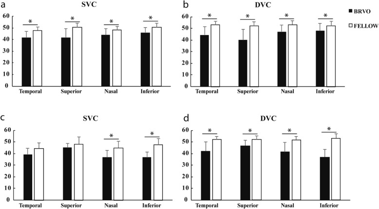
Results: VDs of SCV and DVC were significantly lower, especially in affected regions, in BRVO eyes compared with fellow eyes (P < 0.05). BRVO affected eyes has larger FAZ size, FAZ perimeter, AI and lower FD-300 compared with fellow eyes (all P < 0.05). VD of SVC and FD-300 were lower in fellow eyes compared with normal control eyes (P < 0.05). The average vessel density in whole area and peripapillary area in BRVO eyes were significantly lower compared with fellow eyes (P < 0.05). VD of inside disc in fellow eyes was lower than normal eyes (P < 0.05).
Conclusions: OCTA provided quantitative information of vascular changes in BRVO. FAZ in BRVO eyes showed significant morphological alterations and decreases of VD in surrounding area. Decreases of VD existed not only in SVC and DVC in macular region but also in RPCs in BRVO eyes. Unaffected eyes of unilateral BRVO showed vascular abnormalities in superficial retinal layer, peri-FAZ area and also peripapillary regions.
Keywords: Branch retinal vein occlusion; Foveal avascular zone; Optical coherence tomography angiography; Radial peripapillary capillary; Retinal vasculature.
Conflict of interest statement
The authors declare that they have no competing interests.
Figures

References
MeSH terms
LinkOut - more resources
Full Text Sources
