. 2020 Apr 19;15(1):157.
doi: 10.1186/s13018-020-01665-y.

Characterization of dimensional, morphological and morphometric features of retrieved 3D-printed acetabular cups for hip arthroplasty

Affiliations

Characterization of dimensional, morphological and morphometric features of retrieved 3D-printed acetabular cups for hip arthroplasty

Lorenzo Dall'Ava et al. J Orthop Surg Res. .

Abstract

Background: Three-dimensional (3D) printing of porous titanium implants is increasing in orthopaedics, promising enhanced bony fixation whilst maintaining design similarities with conventionally manufactured components. Our study is one of the first to non-destructively characterize 3D-printed implants, using conventionally manufactured components as a reference.

Methods: We analysed 16 acetabular cups retrieved from patients, divided into two groups: '3D-printed' (n = 6) and 'conventional' (n = 10). Coordinate-measuring machine (CMM), electron microscopy (SEM) and microcomputed tomography (micro-CT) were used to investigate the roundness of the internal cup surface, the morphology of the backside surface and the morphometric features of the porous structures of the cups, respectively. The amount of bony attachment was also evaluated.

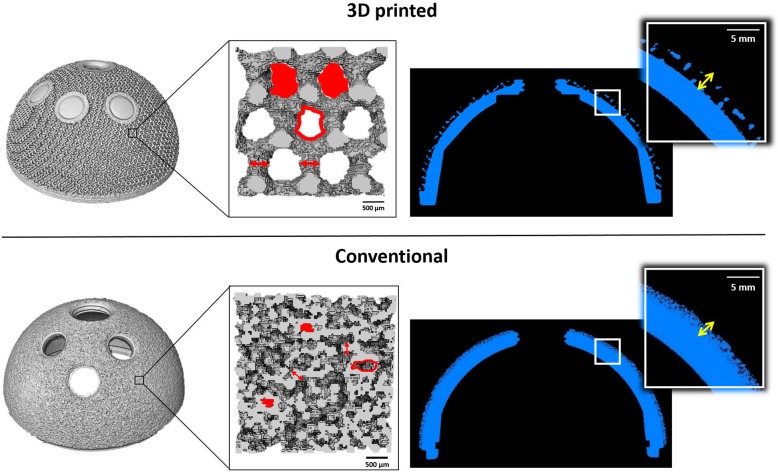
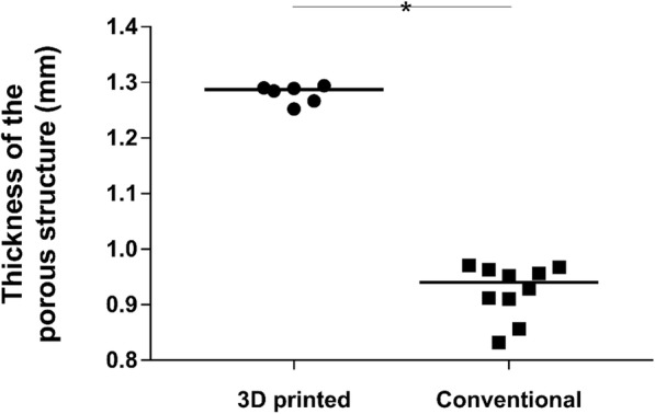
Results: CMM analysis showed a median roundness of 19.45 and 14.52 μm for 3D-printed and conventional cups, respectively (p = 0.1114). SEM images revealed partially molten particles on the struts of 3D-printed implants; these are a by-product of the manufacturing technique, unlike the beads shown by conventional cups. As expected, porosity, pore size, strut thickness and thickness of the porous structure were significantly higher for 3D-printed components (p = 0.0002), with median values of 72.3%, 915 μm, 498 μm and 1.287 mm (p = 0.0002). The median values of bony attachment were 84.9% and 69.3% for 3D-printed and conventional cups, respectively (p = 0.2635).

Conclusion: 3D-printed implants are designed to be significantly more porous than some conventional components, as shown in this study, whilst still exhibiting the same shape and size. We found differences in the surface morphologies of the groups, related to the different manufacturing methods; a key finding was the presence of partially molten particles on the 3D-printed cups.

Keywords: 3D printing; Additive manufacturing; Hip arthroplasty; Orthopaedic implants; Porous acetabular cups.


Conflict of interest statement

The authors declare that they have no competing interests.

Figures

Fig. 1
Fig. 1
Flowchart of the study design
Fig. 2
Fig. 2
Images showing the outcomes of the optimization process of the micro-CT scanning parameters. From top to bottom, the investigated parameters were 80 kV, 200 μA, 100 kV, 100 μA and 150 kV, 70 μA. The set of values 150 kV, 70 μA was chosen because the peak corresponding to the metal (yellow arrow) in the voxel count histogram was higher compared to the same peak in the plots related to the other set of parameters. A higher peak suggested a better separation between voxels belonging to the background (air) and voxels belonging to the material. The reconstructed volume obtained with the values 150 kV, 70 μA provided a clearer representation of the implant compared to the others
Fig. 3
Fig. 3
SEM images of a 3D-printed and a conventional implant. a The 3D-printed porous structure had a regular shape with clearly identifiable pores; b partially molten particles (black circles) were visible on the struts; c the ‘stair-step’ effect due to the layer-over-layer manufacturing and (d) the texture lines were also found at higher magnification. Differently, conventional cups showed e less clearly identifiable pores and f beads of uniform distribution along all the backside surface
Fig. 4
Fig. 4
Images showing the outcomes from micro-CT analysis of 3D-printed (above solid line) and conventional (below solid line) cups. Left to right, the whole reconstructed implants and a zoom on the porous structures with the morphometric parameters depicted (porosity, pore size, strut thickness) are exhibited. Porosity is defined as the percentage of void volume (red filled shapes) over total volume; pore size is the diameter of the circle whose area equals the one of the red empty shapes; and strut thickness is indicated by the red arrows. A cross-section of the components and a zoom showing the thickness of the porous structure (yellow arrows) are also shown. 3D-printed cups showed a more porous and thicker porous backside structures
Fig. 5
Fig. 5
Dot plot showing the distribution of the values of thickness of the porous structure measured for the two groups. The solid line represents the median value. 3D-printed implants showed a significantly thicker structure than conventional cups (p = 0.0002)
Fig. 6
Fig. 6
Image showing a CT cross-section slice of a 3D-printed cup with grey areas (white arrows) depicting presence of a material different from the Ti6Al4V alloy and background air
Fig. 7
Fig. 7
Image showing an example of a low and b high percentage of bony attachment on 3D-printed cups. The presence of bone in the image a is indicated by the yellow arrows

Similar articles

Cited by

References

    1. National Joint Registry for England, Wales NI and I of M. 15th Annual Report [Internet]. 2018 [cited 2019 Apr 1]. Available from: http://www.njrcentre.org.uk.
    1. Murr LE, Gaytan SM, Martinez E, Medina F, Wicker RB. Next generation orthopaedic implants by additive manufacturing using electron beam melting. Int J Biomater. 2012;2012. - PMC - PubMed
    1. Banerjee S, Kulesha G, Kester M, Mont MA. Emerging technologies in arthroplasty: additive manufacturing. J Knee Surg. 2014;27:185–191. doi: 10.1055/s-0034-1374810. - DOI - PubMed
    1. Hart AJ, Hart A, Panagiotopoulou V, Henckel J. Personalised orthopaedics – using 3D printing for tailor-made technical teaching , pre-operative planning and precise placement of implants. Orthop Prod News. 2017;.
    1. Mumith A, Thomas M, Shah Z, Coathup M, Blunn G. Additive manufacturing. Bone Joint J. 2018;100-B:455–460. doi: 10.1302/0301-620X.100B4.BJJ-2017-0662.R2. - DOI - PubMed