. 2020 Nov 1;528(16):2708-2728.
doi: 10.1002/cne.24927. Epub 2020 Jun 11.

Central afferents to the nucleus of the solitary tract in rats and mice

Affiliations

Central afferents to the nucleus of the solitary tract in rats and mice

Silvia Gasparini et al. J Comp Neurol. .

Abstract

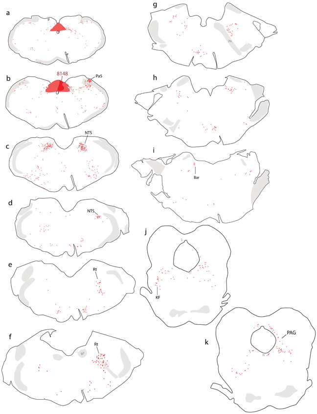
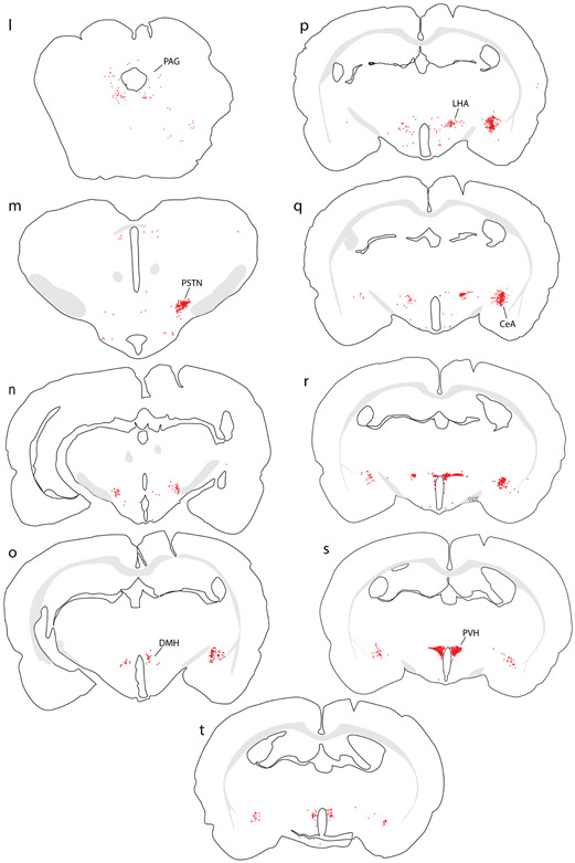
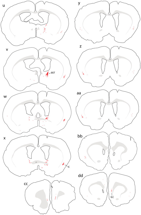
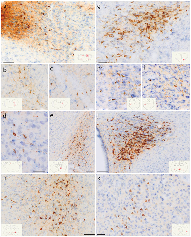
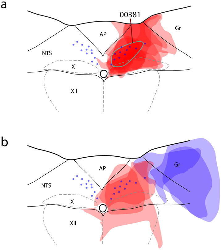
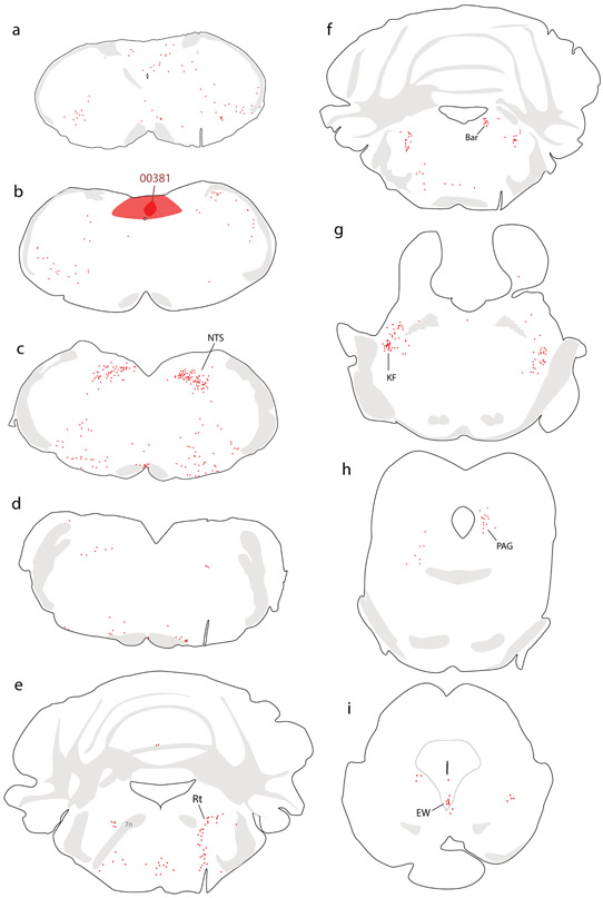
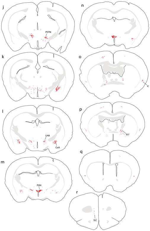
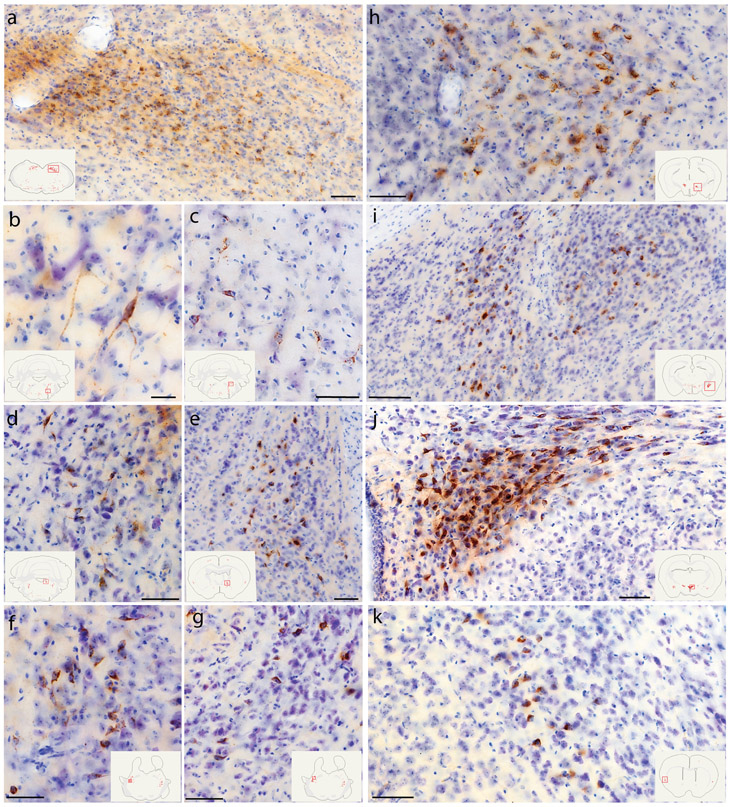
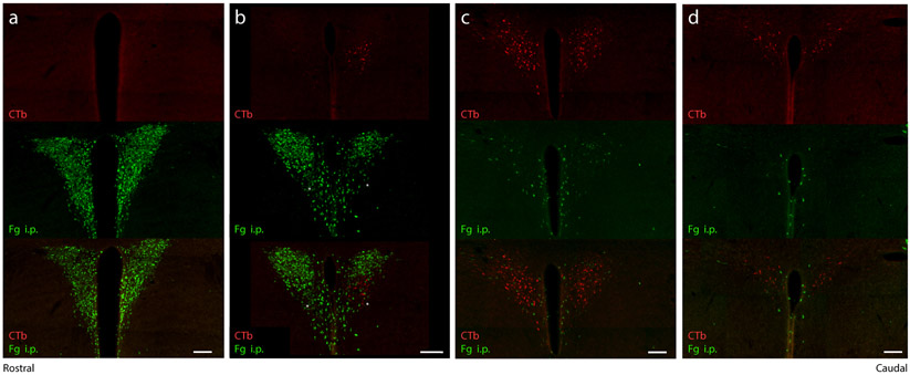
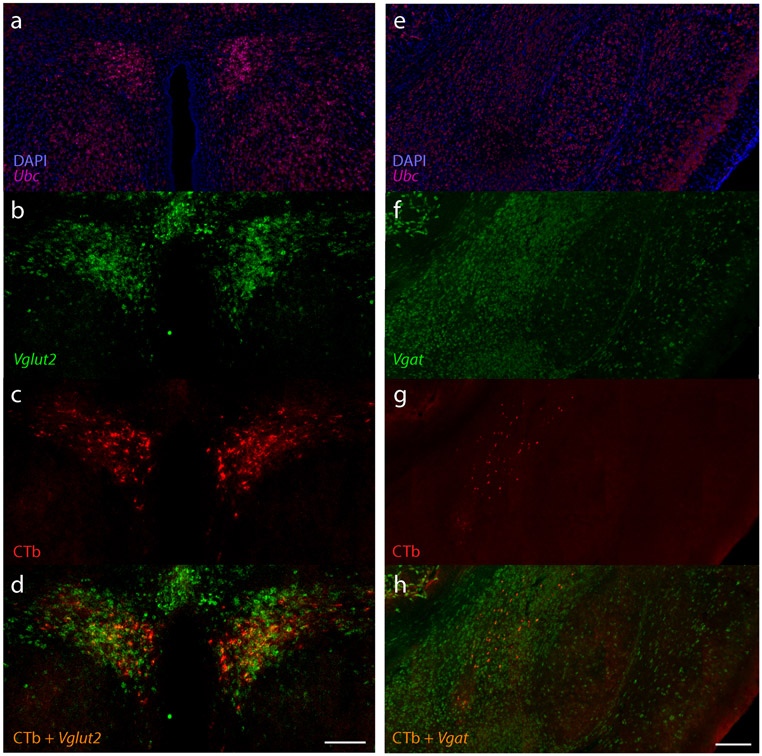
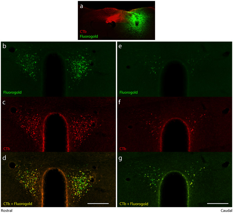
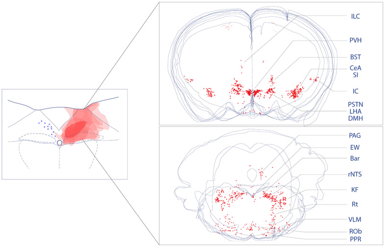
The nucleus of the solitary tract (NTS) regulates life-sustaining functions ranging from appetite and digestion to heart rate and breathing. It is also the brain's primary sensory nucleus for visceral sensations relevant to symptoms in medical and psychiatric disorders. To better understand which neurons may exert top-down control over the NTS, here we provide a brain-wide map of all neurons that project axons directly to the caudal, viscerosensory NTS, focusing on a medial subregion with aldosterone-sensitive HSD2 neurons. Injecting an axonal tracer (cholera toxin b) into the NTS produces a similar pattern of retrograde labeling in rats and mice. The paraventricular hypothalamic nucleus (PVH), lateral hypothalamic area, and central nucleus of the amygdala (CeA) contain the densest concentrations of NTS-projecting neurons. PVH afferents are glutamatergic (express Slc17a6/Vglut2) and are distinct from neuroendocrine PVH neurons. CeA afferents are GABAergic (express Slc32a1/Vgat) and are distributed largely in the medial CeA subdivision. Other retrogradely labeled neurons are located in a variety of brain regions, including the cerebral cortex (insular and infralimbic areas), bed nucleus of the stria terminalis, periaqueductal gray, Barrington's nucleus, Kölliker-Fuse nucleus, hindbrain reticular formation, and rostral NTS. Similar patterns of retrograde labeling result from tracer injections into different NTS subdivisions, with dual retrograde tracing revealing that many afferent neurons project axon collaterals to both the lateral and medial NTS subdivisions. This information provides a roadmap for studying descending axonal projections that may influence visceromotor systems and visceral "mind-body" symptoms.

Keywords: hypothalamus; infralimbic; insular cortex; medial prefrontal cortex; nucleus of the solitary tract; nucleus tractus solitarii; nucleus tractus solitarius; paraventricular; retrograde tracing; solitary nucleus.


Figures

Figure 1.
Figure 1.
Retrograde labeling throughout the brain after cholera toxin B subunit (CTb) injection into the NTS in rat. Abbreviations: 7n, cranial nerve 7 (facial nerve); ac, anterior commissure; Bar, Barrington’s nucleus; BST, bed nucleus of the stria terminalis; CeA, central nucleus of the amygdala; DMH, dorsomedial nucleus of the hypothalamus; g7, genu of cranial nerve 7 (facial nerve); ILC, infralimbic cortex; KF, Kölliker-Fuse nucleus; LHA, lateral hypothalamic area; NTS, nucleus of the solitary tract; opt, optic tract; Pa5, paratrigeminal nucleus; PAG, periaqueductal gray matter; PSTN, parasubthalamic nucleus; PVH, paraventricular nucleus of the hypothalamus; Rt, brainstem reticular formation.
Figure 1.
Figure 1.
Retrograde labeling throughout the brain after cholera toxin B subunit (CTb) injection into the NTS in rat. Abbreviations: 7n, cranial nerve 7 (facial nerve); ac, anterior commissure; Bar, Barrington’s nucleus; BST, bed nucleus of the stria terminalis; CeA, central nucleus of the amygdala; DMH, dorsomedial nucleus of the hypothalamus; g7, genu of cranial nerve 7 (facial nerve); ILC, infralimbic cortex; KF, Kölliker-Fuse nucleus; LHA, lateral hypothalamic area; NTS, nucleus of the solitary tract; opt, optic tract; Pa5, paratrigeminal nucleus; PAG, periaqueductal gray matter; PSTN, parasubthalamic nucleus; PVH, paraventricular nucleus of the hypothalamus; Rt, brainstem reticular formation.
Figure 2.
Figure 2.
CTb (brown) retrograde labeling in select regions of the rat brain, with light Nissl-counterstain (thionin, blue) for cytoarchitectural reference. Inset illustrations highlight the section (plotted in Figure 1) from which each photomicrograph was taken. (a) Rostral NTS; (b-c) hindbrain reticular nucleus along the intramedullary facial nerve fibers; (d) Bar; (e) dorsolateral BST; (f) Medial CeA; (g) PSTN; (h-i) KF region; (j) PVH; (k) mid-insular cortex. Scale bars are 50 μm (b-d, g-k) or 100 μm (a, e, f, j).
Figure 3.
Figure 3.
Injection sites of all mice. (a) CTb injection sites are semi-transparent in red. Blue dots represent the distribution of NTS neurons containing immunoreactivity for 11-beta-hydroxysteroid dehydrogenase type 2 (HSD2), used to align the injection site drawing from each case. The CTb injection site from case #381, used for whole-brain illustration in the next figure, is highlighted with a blue-green outline. Double-retrograde injection cases are shown in (b), with CTb in red and fluorogold (Fg) injection sites in blue semi-transparency. Abbreviations: AP, area postrema; Gr, gracile nucleus; X, dorsal motor nucleus of the vagus nerve; XII, hypoglossal motor nucleus.
Figure 4.
Figure 4.
Retrograde labeling throughout the brain after CTb injection into the NTS in mouse. Additional abbreviations: EW, Edinger-Westphal nucleus.
Figure 4.
Figure 4.
Retrograde labeling throughout the brain after CTb injection into the NTS in mouse. Additional abbreviations: EW, Edinger-Westphal nucleus.
Figure 5.
Figure 5.
CTb (brown) retrograde labeling in select regions of the mouse brain, with light Nissl-counterstain (thionin, blue) for cytoarchitectural reference. In each panel, the inset illustration highlights the region from which each photomicrograph was taken. (a) Rostral NTS; (b-c) hindbrain reticular nucleus; (d) Bar; (e) dorsolateral BST; (f-g) KF region; (h) PSTN; (i) CeA; (j) PVH; (k) mid-insular cortex. All scale bars are 50 μm.
Figure 6.
Figure 6.
Consecutive rostral-to-caudal levels of the PVH (A-D, 1-in-3 40 μm brain sections) showing Fg (green) uptake from systemic (intraperitoneal) injection and CTb retrograde labeling (red) after injection into the NTS. Magnocellular and parvocellular neuroendocrine neurons in the PVH (green) are entirely distinct from (and largely rostral to) PVH neurons that project axons to the NTS (red). Scale bars are 100 μm.
Figure 7.
Figure 7.
(a-d) Expression of mRNA for the vesicular glutamate transporter 2 (Vglut2/Slc17a6, green) shows that CTb retrogradely labeled neurons in the PVH are glutamatergic. (e-h) Expression of mRNA for the vesicular GABA transporter (Vgat/Slc32a1, green) shows that CTb retrogradely labeled neurons in the CeA are GABAergic. In both cases, the ubiquitously expressed Ubc (green) and DAPI nuclear counterstain are shown for background comparison. Scale bars are 200 μm.
Figure 8.
Figure 8.
Double-retrograde labeling from the medial and lateral NTS. (a) CTb injection into the medial NTS (red) with Fg injection targeting the lateral NTS (green) retrogradely labeled (b-g) overlapping and largely co-localized neurons in the PVH. Scale bars are 200 μm.
Figure 9.
Figure 9.
Overall distribution of neurons that project axons to the NTS. To produce these summary images, we overlaid plots of CTb retrograde labeling (red, Figure 3) from the forebrain (top) and brainstem (bottom). CTb-labeled neurons in the motor cortex are faded because anterograde labeling studies from this one site show a lack of projections to the NTS (see text).

References

    1. Allen GV, & Cechetto DF (1992, January 15). Functional and anatomical organization of cardiovascular pressor and depressor sites in the lateral hypothalamic area: I. Descending projections. J Comp Neurol, 315(3), 313–332. 10.1002/cne.903150307 - DOI - PubMed
    1. Altschuler SM, Ferenci DA, Lynn RB, & Miselis RR (1991, February 8). Representation of the cecum in the lateral dorsal motor nucleus of the vagus nerve and commissural subnucleus of the nucleus tractus solitarii in rat. J Comp Neurol, 304(2), 261–274. 10.1002/cne.903040209 - DOI - PubMed
    1. August JT, Nelson DH, & Thorn GW (1958, November). Response of normal subjects to large amounts of aldosterone. J Clin Invest, 37(11), 1549–1555. 10.1172/JCI103747 - DOI - PMC - PubMed
    1. Bailey TW, Hermes SM, Andresen MC, & Aicher SA (2006, November 15). Cranial visceral afferent pathways through the nucleus of the solitary tract to caudal ventrolateral medulla or paraventricular hypothalamus: target-specific synaptic reliability and convergence patterns. J Neurosci, 26(46), 11893–11902. 10.1523/JNEUROSCI.2044-06.2006 - DOI - PMC - PubMed
    1. Banks D, & Harris MC (1987, May). Activation within dorsal medullary nuclei following stimulation in the hypothalamic paraventricular nucleus in rats. Pflugers Arch, 408(6), 619–627. 10.1007/bf00581165 - DOI - PubMed

Publication types

Substances