. 2020 Sep;72(9):1505-1513.
doi: 10.1002/art.41288. Epub 2020 Aug 14.

Attenuation of Murine Collagen-Induced Arthritis by Targeting CD6

Affiliations

Attenuation of Murine Collagen-Induced Arthritis by Targeting CD6

Yan Li et al. Arthritis Rheumatol. 2020 Sep.

Abstract

Objective: CD6 is an important regulator of T cell function that interacts with the ligands CD166 and CD318. To further clarify the significance of CD6 in rheumatoid arthritis (RA), we examined the effects of targeting CD6 in the mouse model of collagen-induced arthritis (CIA), using CD6-knockout (CD6-KO) mice and CD6-humanized mice that express human CD6 in lieu of mouse CD6 on their T cells.

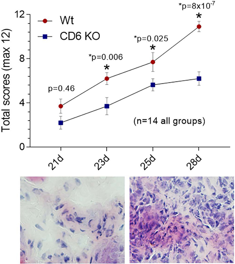
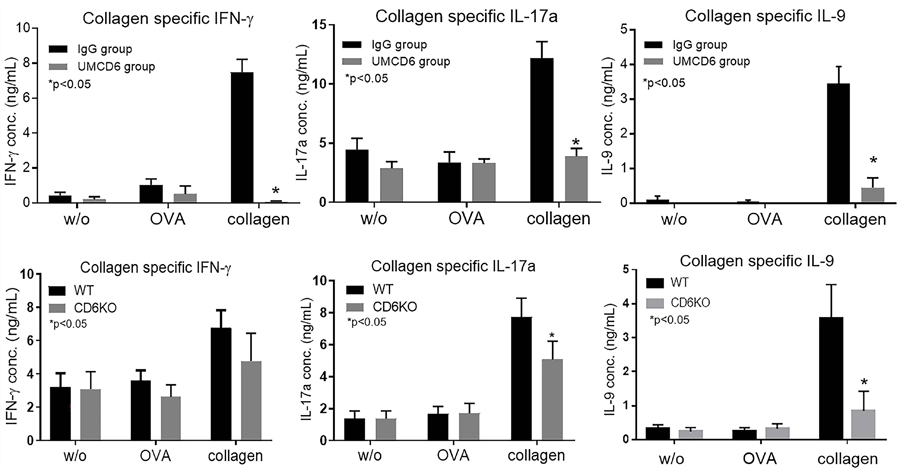
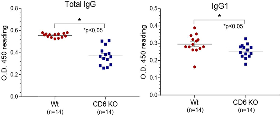
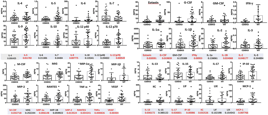
Methods: We immunized wild-type (WT) and CD6 gene-KO mice with a collagen emulsion to induce CIA. For treatment studies using CD6-humanized mice, mice were immunized similarly and a mouse anti-human CD6 IgG (UMCD6) or control IgG was injected on days 7, 14, and 21. Joint tissues were evaluated for tissue damage, leukocyte infiltration, and local inflammatory cytokine production. Collagen-specific Th1, Th9, and Th17 responses and serum levels of collagen-specific IgG subclasses were also evaluated in WT and CD6-KO mice with CIA.

Results: The absence of CD6 reduced 1) collagen-specific Th9 and Th17, but not Th1 responses, 2) the levels of many proinflammatory joint cytokines, and 3) serum levels of collagen-reactive total IgG and IgG1, but not IgG2a and IgG3. Joint homogenate hemoglobin content was significantly reduced in CD6-KO mice with CIA compared to WT mice with CIA (P < 0.05) (reduced angiogenesis). Moreover, treating CD6-humanized mice with mouse anti-human CD6 monoclonal antibody was similarly effective in reducing joint inflammation in CIA.

Conclusion: Taken together, these data suggest that interaction of CD6 with its ligands is important for the perpetuation of CIA and other inflammatory arthritides that are T cell driven.


Conflict of interest statement

Conflicts of interest: none

Figures

Figure 1.
Figure 1.. Clinical scoring shows reduction in CIA severity in CD6 knockout (KO) mice compared to Wt mice.
CIA severity was measured in each paw and ankle (front and hind joints). A total reduction in CIA severity was evident on days 23 and beyond. On day 28, Wt mice have a robust synovial inflammatory response (lower right panel) that is not evident in the CD6 KO mice (lower left panel, n=14 in each group). Data are presented as mean ± SEM (*p<0.05).
Figure 2.
Figure 2.. Clinical scoring shows a reduction of CIA severity in CD6-humanized mice administered UMCD6.
CIA severity was measured in each paw and ankle (front and hind joints) from CD6-humanized mice with CIA treated with either human IgG or UMCD6 antibody on days 7, 14 and 21. A total reduction of CIA severity was evident on days 28 and beyond. The lower left panel shows that mice administered IgG have demonstrable synovial CD3+ lymphocyte staining (fluorescent green cells) on day 35, that is not evident in the UMCD6 treated mice (lower right panel, n=14 in each group) data were mean ± SEM (*p<0.05).
Figure 3.
Figure 3.. Reduced T cell cytokine production in response to collagen due to blocking or absence of CD6.
Th1//Th17/Th9 recall assays in UMCD6 treatment studies show that splenocytes from the IgG treated CIA group, but not the UMCD6 treated CIA mice, showed reactivity to collagen in vitro as measured by secretion of IFN-γ, IL-17 and IL-9. Controls include no antigen and a non-relevant antigen (OVA 323–339 peptide) (upper panel). We also performed collagen-specific recall assays using splenocytes collected from CD6 KO or Wt mice after CIA induction. These assays demonstrated significantly reduced collagen-specific Th17 and Th9 but not Th1 responses (lower panel). The ELISA assays have sensitivities in the picogram range for each of the cytokines tested. Data are mean ± SEM (*p<0.05).
Figure 4.
Figure 4.. Total serum levels of IgG and IgG1 against collagen II are lower in CD6 KO CIA mice.
Total serum levels of IgG and IgG1 anti-collagen II were measured by ELISA in Wt and CD6 KO mice with CIA. Left panel: Total IgG anti-collagen II, Right panel: anti-collagen II IgG1. Data are mean ± SEM (*p<0.05).
Figure 5.
Figure 5.. Cytokine panels from joint homogenates of CIA CD6 KO and Wt mice.
CIA mouse joints from Wt and CD6 KO mice were homogenized and measured for protein concentrations. A cytokine multiplex assay was used to measure the levels of 32 cytokines in homogenates of each of the paw and ankle joint tissues. Cytokine levels were normalized to protein concentration for each joint tissue homogenate (pg cytokine/mg protein). Many pro-inflammatory cytokines were reduced in CD6 KO CIA mice compared to Wt CIA mice. The numbers listed below each cytokine are p-values for each cytokine measured. Numbers in red indicate p-values<0.05 (statistically significant).

Similar articles

  • CD69 targeting differentially affects the course of collagen-induced arthritis.
    Sancho D, Gómez M, Martinez Del Hoyo G, Lamana A, Esplugues E, Lauzurica P, Martinez-A C, Sánchez-Madrid F. Sancho D, et al. J Leukoc Biol. 2006 Dec;80(6):1233-41. doi: 10.1189/jlb.1205749. Epub 2006 Aug 18. J Leukoc Biol. 2006. PMID: 16921025
  • Targeting CD6 for the treatment of experimental autoimmune uveitis.
    Zhang L, Li Y, Qiu W, Bell BA, Dvorina N, Baldwin WM 3rd, Singer N, Kern T, Caspi RR, Fox DA, Lin F. Zhang L, et al. J Autoimmun. 2018 Jun;90:84-93. doi: 10.1016/j.jaut.2018.02.004. Epub 2018 Feb 19. J Autoimmun. 2018. PMID: 29472120 Free PMC article.
  • CD318 is a ligand for CD6.
    Enyindah-Asonye G, Li Y, Ruth JH, Spassov DS, Hebron KE, Zijlstra A, Moasser MM, Wang B, Singer NG, Cui H, Ohara RA, Rasmussen SM, Fox DA, Lin F. Enyindah-Asonye G, et al. Proc Natl Acad Sci U S A. 2017 Aug 15;114(33):E6912-E6921. doi: 10.1073/pnas.1704008114. Epub 2017 Jul 31. Proc Natl Acad Sci U S A. 2017. PMID: 28760953 Free PMC article.
  • [Animal models for bone and joint disease. CIA, CAIA model].
    Hirose J, Tanaka S. Hirose J, et al. Clin Calcium. 2011 Feb;21(2):253-9. Clin Calcium. 2011. PMID: 21289422 Review. Japanese.
  • CD6 in Human Disease.
    Gurrea-Rubio M, Fox DA, Castresana JS. Gurrea-Rubio M, et al. Cells. 2025 Feb 13;14(4):272. doi: 10.3390/cells14040272. Cells. 2025. PMID: 39996744 Free PMC article. Review.

Cited by

References

    1. Gangemi RM, Swack JA, Gaviria DM, Romain PL. Anti-T12, an anti-CD6 monoclonal antibody, can activate human T lymphocytes. J Immunol. 1989;143(8):2439–47. - PubMed
    1. Consuegra-Fernandez M, Lin F, Fox DA, Lozano F. Clinical and experimental evidence for targeting CD6 in immune-based disorders. Autoimmun Rev. 2018;17(5):493–503. - PubMed
    1. Osorio LM, Ordonez C, Garcia CA, Jondal M, Chow SC. Evidence for protein tyrosine kinase involvement in CD6-induced T cell proliferation. Cell Immunol. 1995;166(1):44–52. - PubMed
    1. Breuning J, Brown MH. T Cell Costimulation by CD6 Is Dependent on Bivalent Binding of a GADS/SLP-76 Complex. Mol Cell Biol. 2017;37(11). - PMC - PubMed
    1. Hem CD, Ekornhol M, Granum S, Sundvold-Gjerstad V, Spurkland A. CD6 and Linker of Activated T Cells are Potential Interaction Partners for T Cell-Specific Adaptor Protein. Scand J Immunol. 2017;85(2):104–12. - PubMed

Publication types

MeSH terms