

Polarizable Molecular Dynamics Simulations of Two c-kit Oncogene Promoter G-Quadruplexes: Effect of Primary and Secondary Structure on Loop and Ion Sampling
- PMID: 32307997
- PMCID: PMC7221321
- DOI: 10.1021/acs.jctc.0c00191
Polarizable Molecular Dynamics Simulations of Two c-kit Oncogene Promoter G-Quadruplexes: Effect of Primary and Secondary Structure on Loop and Ion Sampling
Abstract
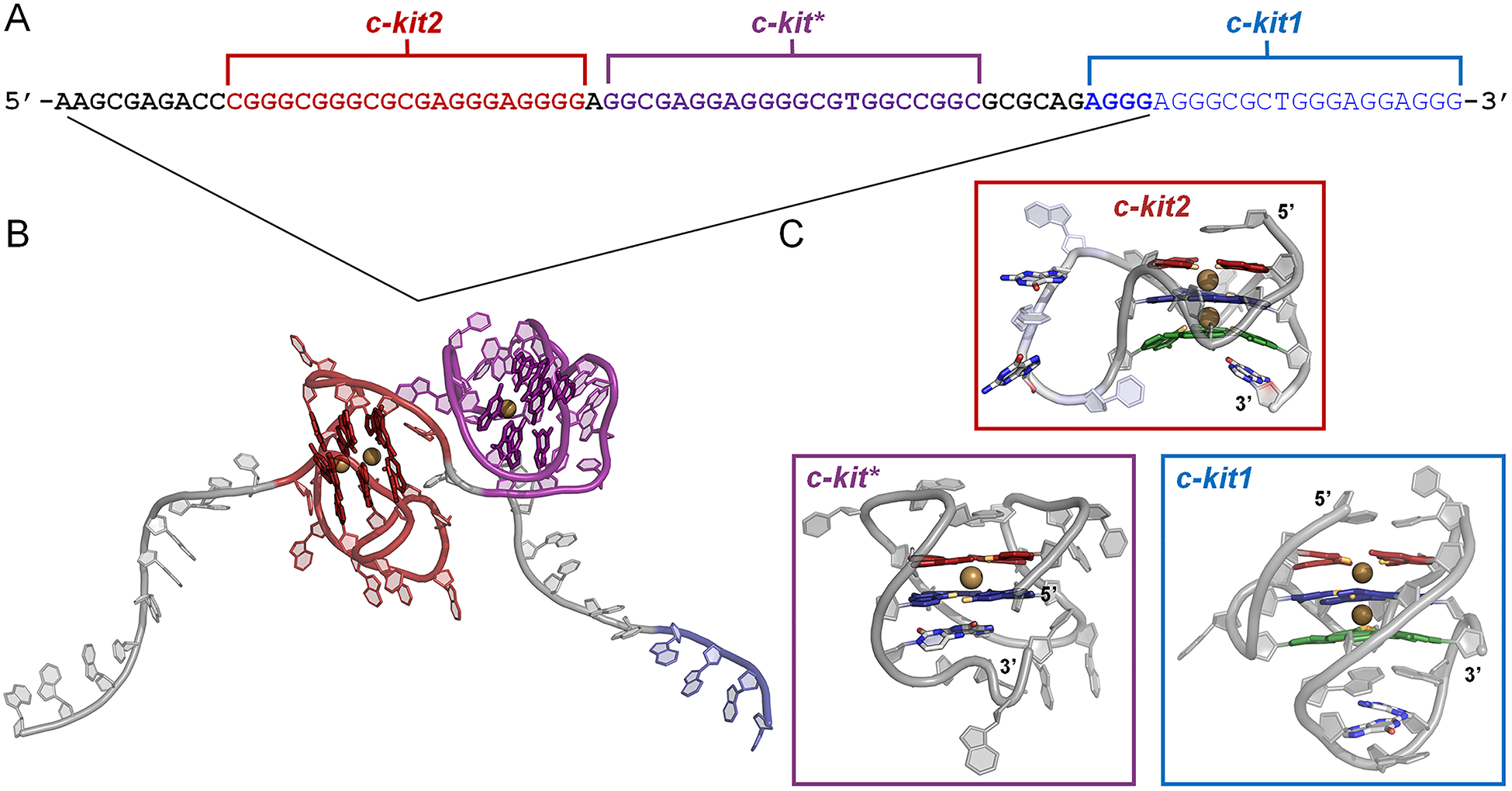
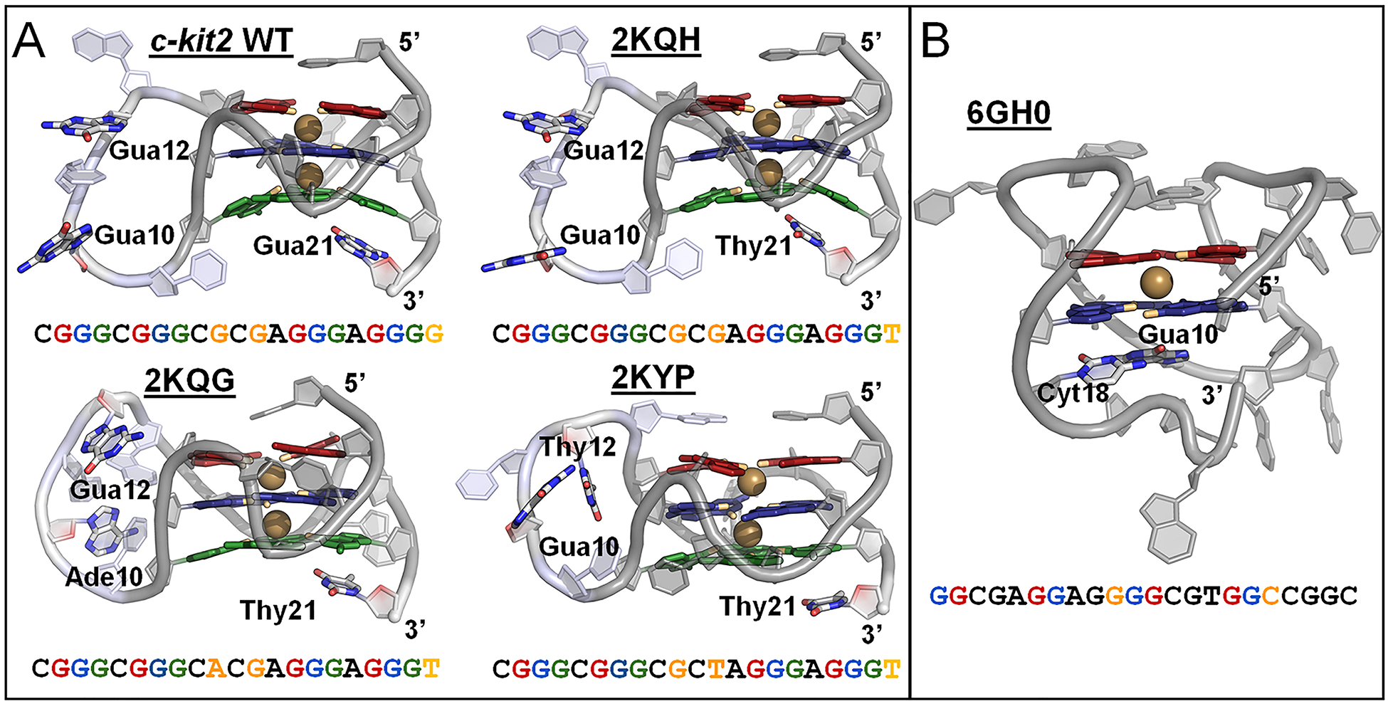
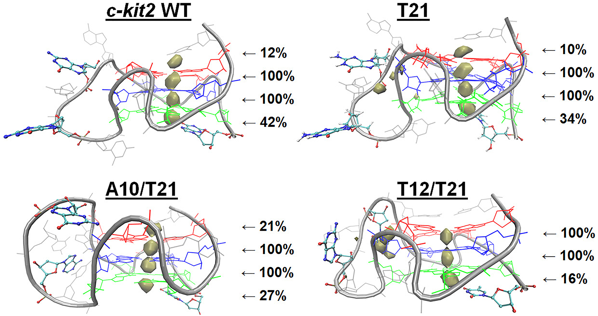
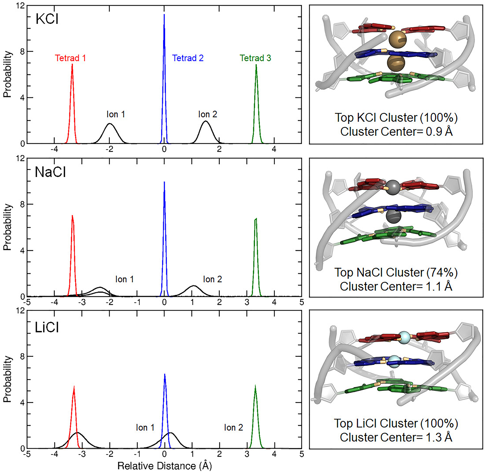
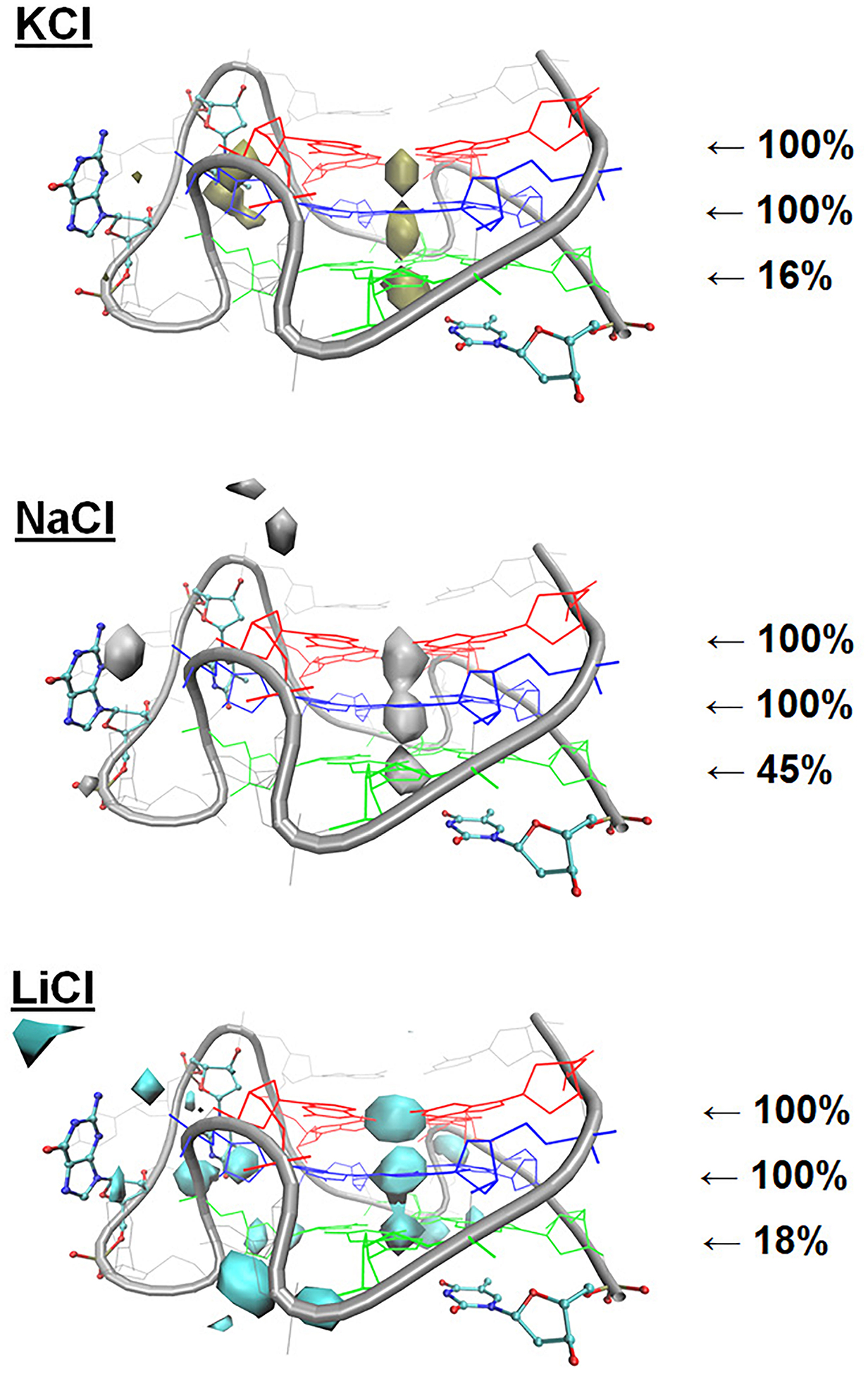
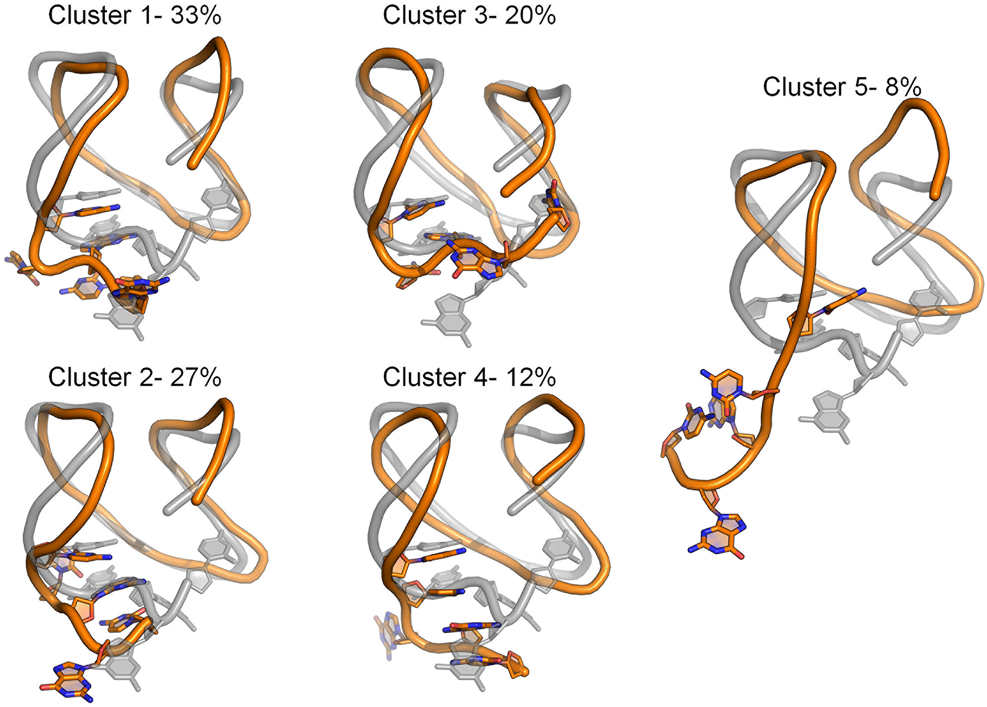
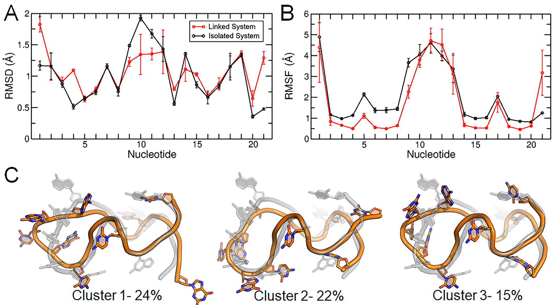
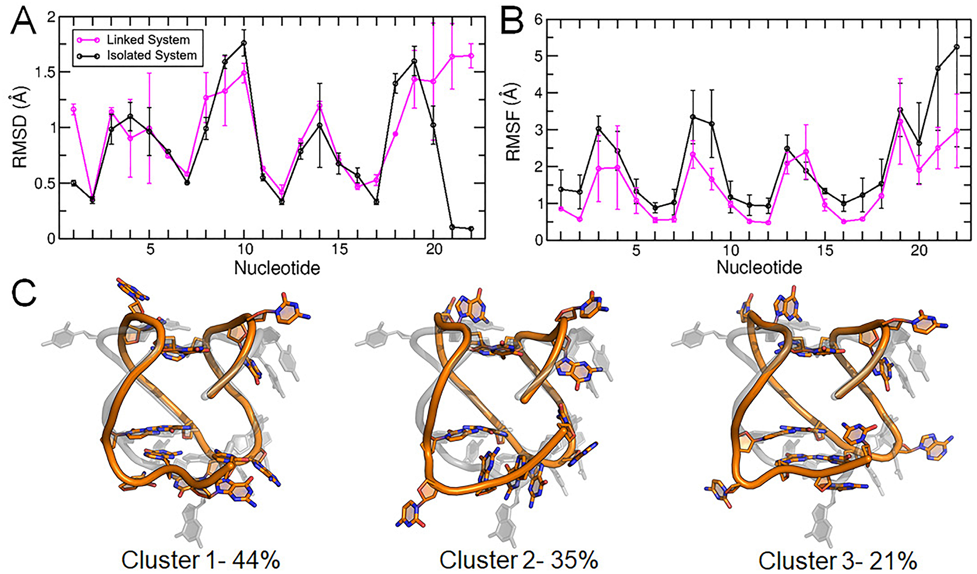
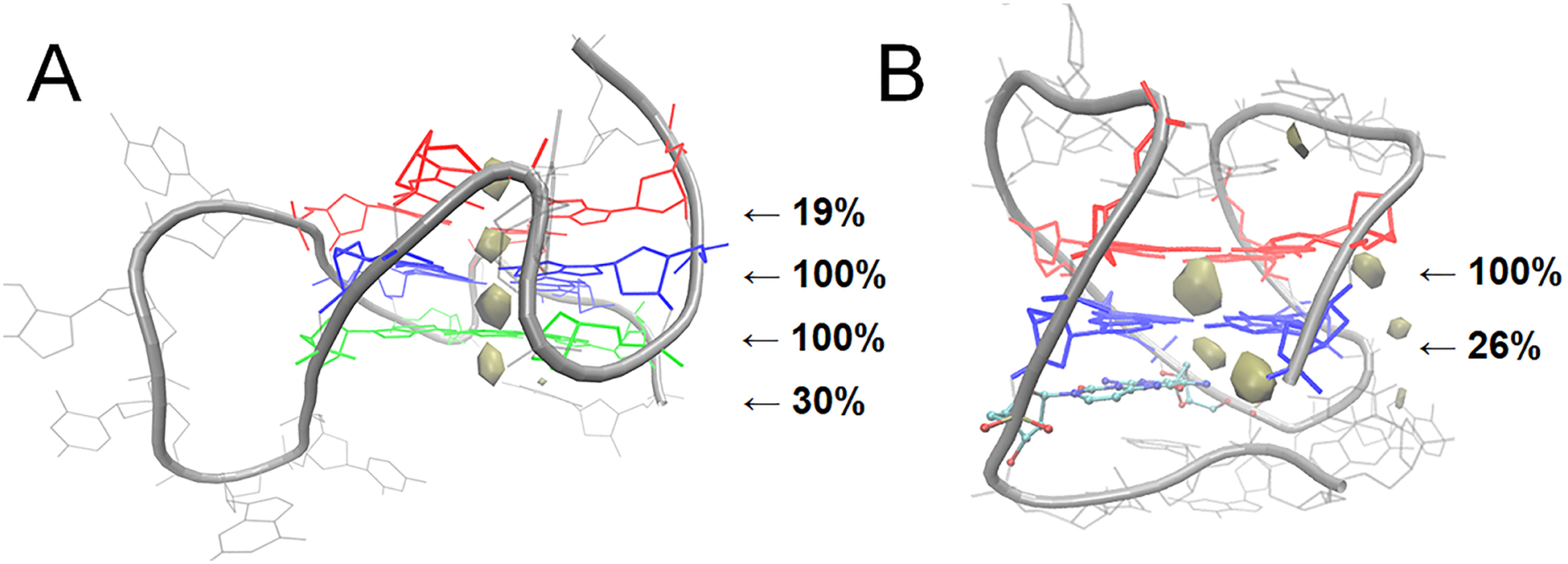
G-quadruplexes (GQs) are highly ordered nucleic acid structures that play fundamental roles in regulating gene expression and maintaining genomic stability. GQs are topologically diverse and enriched in promoter sequences of growth regulatory genes and proto-oncogenes, suggesting that they may serve as attractive targets for drug design at the level of transcription rather than inhibiting the activity of the protein products of these genes. The c-kit promoter contains three adjacent GQ-forming sequences that have proposed antagonistic effects on gene expression and thus are promising drug targets for diseases such as gastrointestinal stromal tumors, mast cell disease, and leukemia. Because GQ stability is influenced by primary structure, secondary structure, and ion interactions, a greater understanding of GQ structure, dynamics, and ion binding properties is needed to develop novel, GQ-targeting therapeutics. Here, we performed molecular dynamics simulations to systematically study the c-kit2 and c-kit* GQs, evaluating nonpolarizable and polarizable force fields (FFs) and examining the effects of base substitutions and cation type (K+, Na+, and Li+) on the dynamics of their isolated and linked structures. We found that the Drude polarizable FF outperformed the additive CHARMM36 FF in two- and three-tetrad GQs and solutions of KCl, NaCl, and LiCl. Drude simulations with different cations agreed with the known GQ stabilization preference (K+ > Na+ > Li+) and illustrated that tetrad core-ion coordination differs as a function of cation type. Finally, we showed that differences in primary and secondary structure influence loop sampling, ion binding, and core-ion energetics of GQs.
Figures
Similar articles
-
Cation competition and recruitment around the c-kit1 G-quadruplex using polarizable simulations.Biophys J. 2021 Jun 1;120(11):2249-2261. doi: 10.1016/j.bpj.2021.03.022. Epub 2021 Mar 29. Biophys J. 2021. PMID: 33794153 Free PMC article.
-
Molecular Dynamics Simulations of the c-kit1 Promoter G-Quadruplex: Importance of Electronic Polarization on Stability and Cooperative Ion Binding.J Phys Chem B. 2019 Jan 10;123(1):148-159. doi: 10.1021/acs.jpcb.8b11026. Epub 2018 Dec 20. J Phys Chem B. 2019. PMID: 30525627
-
Same fold, different properties: polarizable molecular dynamics simulations of telomeric and TERRA G-quadruplexes.Nucleic Acids Res. 2020 Jan 24;48(2):561-575. doi: 10.1093/nar/gkz1154. Nucleic Acids Res. 2020. PMID: 31807754 Free PMC article.
-
Folding of guanine quadruplex molecules-funnel-like mechanism or kinetic partitioning? An overview from MD simulation studies.Biochim Biophys Acta Gen Subj. 2017 May;1861(5 Pt B):1246-1263. doi: 10.1016/j.bbagen.2016.12.008. Epub 2016 Dec 13. Biochim Biophys Acta Gen Subj. 2017. PMID: 27979677 Review.
-
G-quadruplex dynamics.Biochim Biophys Acta Proteins Proteom. 2017 Nov;1865(11 Pt B):1544-1554. doi: 10.1016/j.bbapap.2017.06.012. Epub 2017 Jun 20. Biochim Biophys Acta Proteins Proteom. 2017. PMID: 28642152 Review.
Cited by
-
Nanomechanics of G-quadruplexes within the promoter of the KIT oncogene.Nucleic Acids Res. 2021 May 7;49(8):4564-4573. doi: 10.1093/nar/gkab079. Nucleic Acids Res. 2021. PMID: 33849064 Free PMC article.
-
Atomistic Polarizable Embeddings: Energy, Dynamics, Spectroscopy, and Reactivity.Acc Chem Res. 2021 Jul 6;54(13):2812-2822. doi: 10.1021/acs.accounts.0c00662. Epub 2021 May 7. Acc Chem Res. 2021. PMID: 33961401 Free PMC article.
-
Quantifying Induced Dipole Effects in Small Molecule Permeation in a Model Phospholipid Bilayer.J Phys Chem B. 2024 Aug 1;128(30):7385-7400. doi: 10.1021/acs.jpcb.4c01634. Epub 2024 Jul 22. J Phys Chem B. 2024. PMID: 39038441 Free PMC article.
-
Double-stranded flanking ends affect the folding kinetics and conformational equilibrium of G-quadruplexes forming sequences within the promoter of KIT oncogene.Nucleic Acids Res. 2021 Sep 27;49(17):9724-9737. doi: 10.1093/nar/gkab674. Nucleic Acids Res. 2021. PMID: 34478543 Free PMC article.
-
Base pair dynamics, electrostatics, and thermodynamics at the LTR-III quadruplex:duplex junction.Biophys J. 2024 May 7;123(9):1129-1138. doi: 10.1016/j.bpj.2024.03.042. Epub 2024 Apr 4. Biophys J. 2024. PMID: 38576161 Free PMC article.
References
-
- Mulholland K; Wu C Binding of Telomestatin to a Telomeric G-Quadruplex DNA Probed by All-Atom Molecular Dynamics Simulations with Explicit Solvent. J. Chem. Inf. Model 2016, 56 (10), 2093–2102. - PubMed
MeSH terms
Substances
Grants and funding
LinkOut - more resources
Full Text Sources
Other Literature Sources