. 2020 May 1;318(5):F1306-F1312.
doi: 10.1152/ajprenal.00070.2020. Epub 2020 Apr 20.

Integrin-β1 is required for the renal cystogenesis caused by ciliary defects

Affiliations

Integrin-β1 is required for the renal cystogenesis caused by ciliary defects

Miran Yoo et al. Am J Physiol Renal Physiol. .

Abstract

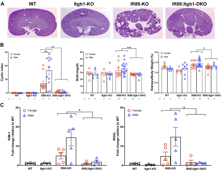
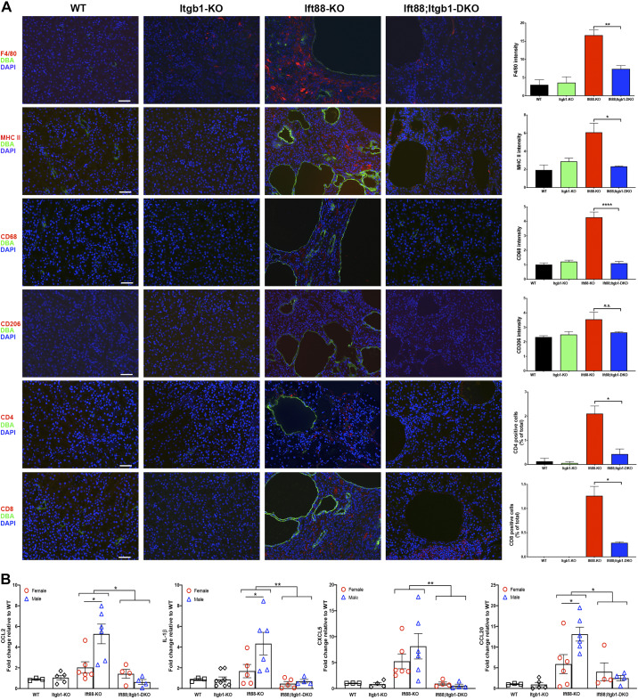
Defects in the function of primary cilia are commonly associated with the development of renal cysts. On the other hand, the intact cilium appears to contribute a cystogenic signal whose effectors remain unclear. As integrin-β1 is required for the cystogenesis caused by the deletion of the polycystin 1 gene, we asked whether it would be similarly important in the cystogenetic process caused by other ciliary defects. We addressed this question by investigating the effect of integrin-β1 deletion in a ciliopathy genetic model in which the Ift88 gene, a component of complex B of intraflagellar transport that is required for the proper assembly of cilia, is specifically ablated in principal cells of the collecting ducts. We showed that the renal cystogenesis caused by loss of Ift88 is prevented when integrin-β1 is simultaneously depleted. In parallel, pathogenetic manifestations of the disease, such as increased inflammatory infiltrate and fibrosis, were also significantly reduced. Overall, our data indicate that integrin-β1 is also required for the renal cystogenesis caused by ciliary defects and point to integrin-β1-controlled pathways as common drivers of the disease and as possible targets to interfere with the cystogenesis caused by ciliary defects.

Keywords: Ift88; ciliopathy; cilium; cystic kidney; integrin.


Conflict of interest statement

None of the authors has any conflicts of interest, financial or otherwise, to disclose.

Figures

Fig. 1.
Fig. 1.
Integrin-β1 (Itgb1 gene) deletion attenuates renal cystogenesis in Ift88 mutants. A: representative images of 9-mo-old wild-type (WT), Itgb1 knockout (KO), Ift88 KO, and Ift88;Itgb1 double-KO (DKO) kidneys stained with periodic acid-Schiff. B: cystic index (left), blood urea nitrogen (BUN) levels (middle), and kidney-to-body weight ratio (right) in 9-mo-old male and female WT, Itgb1 KO, Ift88 KO, and Ift88;Itgb1 DKO mice [the number of animals in each group (n) is indicated at the bottom of each graph]. C: whole kidney expression of kidney injury molecule-1 (KIM-1) and neutrophil gelatinase-associated lipocalin (NGAL) as determined by quantitative RT-PCR. *P < 0.05; **P < 0.01; ***P < 0.001.
Fig. 2.
Fig. 2.
Inactivation of integrin-β1 (Itgb1 gene) reduced fibrosis in Ift88 mutants. A, top row: representative immunofluorescence images of wild-type (WT; n = 3), Itgb1 knockout (KO; n = 3), Ift88 KO (n = 3), and Ift88;Itgb1 double-KO (DKO) kidneys (n = 3) stained with Ki-67 (green), Dolichos biflorus agglutinin (DBA; red), and DAPI (blue). Scale bar = 50 μm. The graph on the right shows the quantification of Ki-67-positive cells relative to total cell number (5 fields/mouse). A, bottom row: representative TUNEL images of WT (n = 4), Itgb1 KO (n = 4), Ift88 KO (n = 4), and Ift88;Itgb1 DKO (n = 4) kidneys. Scale bar = 50 μm. The graph on the right shows the corresponding quantification of positive cells relative to total cells (5 fields/mouse). B, top row: representative staining for hematoxylin and eosin of WT, Itgb1 KO, Ift88 KO, and Ift88;Itgb1 DKO kidneys showing the abundant infiltrate. B, middle row: sirius red staining with relative quantification of the positive area from whole kidney sections from WT (n = 2), Itgb1 KO (n = 4), Ift88 KO (n = 6), and Ift88;Itgb1 DKO (n = 5) mice (5 fields/mouse). B, bottom row: representative immunofluorescence images of WT (n = 2), Itgb1 KO (n = 3), Ift88 KO (n = 3), and Ift88;Itgb1 DKO (n = 2) kidneys stained with α-smooth muscle actin (α-SMA; red) and DBA (green) and counterstained with DAPI (blue). Scale bar = 50 μm. The graph on the right shows relative fluorescence intensity measurements (5 fields/mouse). C: whole kidney expression of fibronectin, collagen type I, and tissue inhibitor of metalloproteinase-1 (Timp1) mRNAs as determined. All kidneys were from 9-mo-old animals. *P < 0.05; **P < 0.01; ***P = 0.0001; ****P < 0.0001.
Fig. 3.
Fig. 3.
Inactivation of integrin-β1 (Itgb1 gene) reduces the inflammation in the kidneys of Ift88 mutants. A: representative immunofluorescence images of wild-type (WT), Itgb1 knockout (KO), Ift88 KO, and Ift88;Itgb1 double-KO (DKO) kidneys costained for Dolichos biflorus agglutinin (DBA; green) and F4/80 (red, first row), major histocompatibility complex class II (MHC II; red, second row), CD68 (red, third row), CD206 (red, fourth row), CD4 (red, fifth row), or CD8 (red, sixth row) and then counterstained with DAPI (blue). Scale bars = 50 μm. The graphs on the right show the corresponding fluorescence intensities determined from n = 2–5/group (5 fields/mouse). B: whole kidney expression of chemokine (C-C motif) ligand (CCL)2, IL1-β, chemokine (C-X-C motif) ligand (CXCL)5, and CCL20 mRNAs as determined by quantitative RT-PCR. All kidneys were from 9-mo-old animals. *P < 0.05; **P < 0.01.

References

    1. Anvarian Z, Mykytyn K, Mukhopadhyay S, Pedersen LB, Christensen ST. Cellular signalling by primary cilia in development, organ function and disease. Nat Rev Nephrol 15: 199–219, 2019. doi:10.1038/s41581-019-0116-9. - DOI - PMC - PubMed
    1. Attanasio M, Uhlenhaut NH, Sousa VH, O’Toole JF, Otto E, Anlag K, Klugmann C, Treier A-C, Helou J, Sayer JA, Seelow D, Nürnberg G, Becker C, Chudley AE, Nürnberg P, Hildebrandt F, Treier M. Loss of GLIS2 causes nephronophthisis in humans and mice by increased apoptosis and fibrosis. Nat Genet 39: 1018–1024, 2007. doi:10.1038/ng2072. - DOI - PubMed
    1. Bell PD, Fitzgibbon W, Sas K, Stenbit AE, Amria M, Houston A, Reichert R, Gilley S, Siegal GP, Bissler J, Bilgen M, Chou PC-T, Guay-Woodford L, Yoder B, Haycraft CJ, Siroky B. Loss of primary cilia upregulates renal hypertrophic signaling and promotes cystogenesis. J Am Soc Nephrol 22: 839–848, 2011. doi:10.1681/ASN.2010050526. - DOI - PMC - PubMed
    1. Berbari NF, Sharma N, Malarkey EB, Pieczynski JN, Boddu R, Gaertig J, Guay-Woodford L, Yoder BK. Microtubule modifications and stability are altered by cilia perturbation and in cystic kidney disease. Cytoskeleton (Hoboken) 70: 24–31, 2013. doi:10.1002/cm.21088. - DOI - PMC - PubMed
    1. Blitzer AL, Panagis L, Gusella GL, Danias J, Mlodzik M, Iomini C. Primary cilia dynamics instruct tissue patterning and repair of corneal endothelium. Proc Natl Acad Sci USA 108: 2819–2824, 2011. doi:10.1073/pnas.1016702108. - DOI - PMC - PubMed

Publication types

MeSH terms