

LncRNA-GAS5 regulates PDCD4 expression and mediates myocardial infarction-induced cardiomyocytes apoptosis via targeting MiR-21
- PMID: 32308118
- PMCID: PMC7469558
- DOI: 10.1080/15384101.2020.1750257
LncRNA-GAS5 regulates PDCD4 expression and mediates myocardial infarction-induced cardiomyocytes apoptosis via targeting MiR-21
Abstract
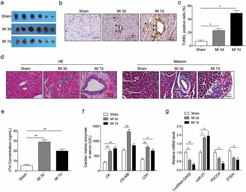
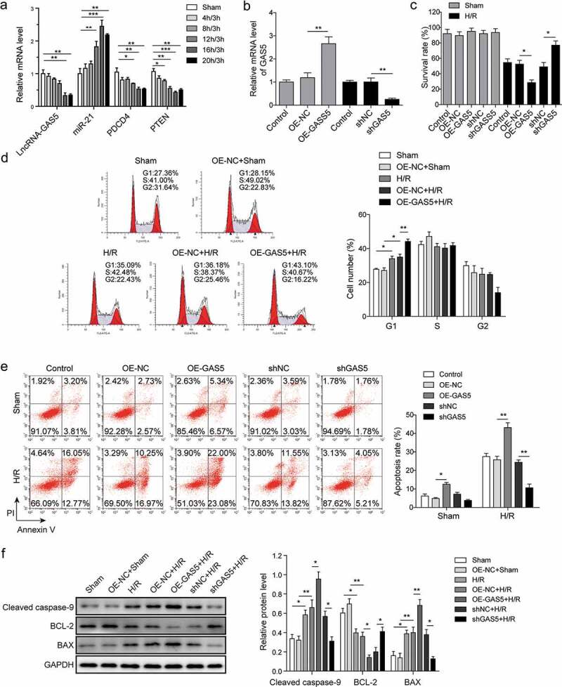
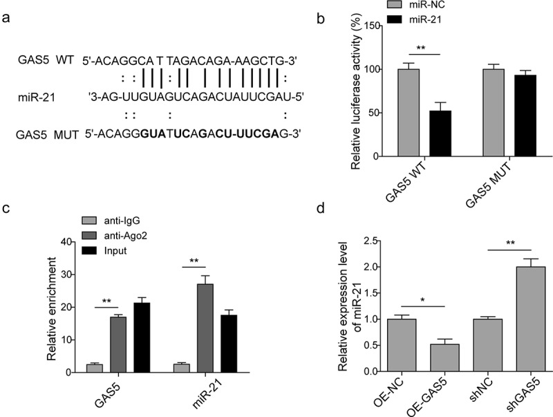
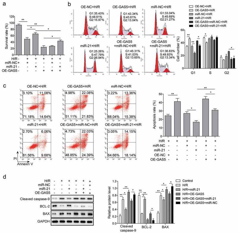
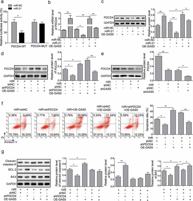
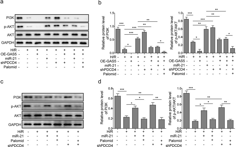
The present study was designed to investigate whether and how lncRNA-GAS5 regulates cardiomyocyte apoptosis in MI. MI rat model was established by the left anterior descending (LAD) coronary artery ligation. MI model was further evaluated by biomarkers detection and TUNEL, HE and Masson staining. The roles of lncRNA-GAS5 on hypoxia/reoxygenation (H/R)-induced cardiomyocytes survival, cell cycle arrest, and apoptosis were examined by MTT and flow cytometry in rat heart-derived H9c2 cells. Western blot was used to determine the effect of GAS5 on the expression of apoptosis-associated proteins and PI3 K/AKT signaling pathway. The direct bindings of GAS5 to miR-21 and miR-21 to PDCD4 were measured by dual-luciferase reporter assay or RNA immunoprecipitation. Decreased expressions of GAS5 and PDCD4 as well as increased miR-21 level were observed in the hearts of MI-modeled rat, accompanying with morphologically myocardial cell injury, as well as collagen deposition and fibrosis, and elevated levels of cTnl, CK, CK-MB and LDH. In the cell model, the knockdown of GAS5 promoted cell survival, prevented cell cycle arrest and inhibited cell apoptosis while the overexpression of GAS5 showed the opposite effects. GAS5 was found to downregulate miR-21 and the effects of GAS5 were attenuated by miR-21 mimics. GAS5 positively regulated PDCD4 expression by functioning as a sponge of miR-21 in H/R model. Moreover, GAS5 stimulated PDCD4 and suppressed PI3 K/AKT signal pathway. LncRNA-GAS5 regulates PDCD4 expression to mediate MI-induced cardiomyocyte apoptosis via targeting miR-21, suggesting that GAS5 could be a therapeutic target for MI.
Keywords: Myocardial infarction; PDCD4; apoptosis; lncRNA-GAS5; miR-21.
Conflict of interest statement
The authors declare that they have no conflict of interest.
Figures
Similar articles
-
lncRNA GAS5 regulates myocardial infarction by targeting the miR-525-5p/CALM2 axis.J Cell Biochem. 2019 Nov;120(11):18678-18688. doi: 10.1002/jcb.29156. Epub 2019 Aug 19. J Cell Biochem. 2019. PMID: 31429119
-
Down-regulation of GAS5 ameliorates myocardial ischaemia/reperfusion injury via the miR-335/ROCK1/AKT/GSK-3β axis.J Cell Mol Med. 2019 Dec;23(12):8420-8431. doi: 10.1111/jcmm.14724. Epub 2019 Oct 18. J Cell Mol Med. 2019. PMID: 31625671 Free PMC article.
-
lncRNA HOTAIR Protects Myocardial Infarction Rat by Sponging miR-519d-3p.J Cardiovasc Transl Res. 2019 Jun;12(3):171-183. doi: 10.1007/s12265-018-9839-4. Epub 2019 Jan 3. J Cardiovasc Transl Res. 2019. PMID: 30607799
-
Revisiting miRNA-21 as a Therapeutic Strategy for Myocardial Infarction: A Systematic Review.J Cardiovasc Pharmacol. 2022 Sep 1;80(3):393-406. doi: 10.1097/FJC.0000000000001305. J Cardiovasc Pharmacol. 2022. PMID: 35767710
-
Progress on the relationship between tumor suppressor PDCD4 and diseases based on the analysis of structural characteristics.Yi Chuan. 2024 Apr 20;46(4):290-305. doi: 10.16288/j.yczz.23-291. Yi Chuan. 2024. PMID: 38632092 Review.
Cited by
-
Bone marrow mesenchymal stem cells-derived exosomal lncRNA GAS5 mitigates heart failure by inhibiting UL3/Hippo pathway-mediated ferroptosis.Eur J Med Res. 2024 May 30;29(1):303. doi: 10.1186/s40001-024-01880-x. Eur J Med Res. 2024. PMID: 38812041 Free PMC article.
-
Long Non-coding RNA Involved in the Pathophysiology of Atrial Fibrillation.Cardiovasc Drugs Ther. 2025 Apr;39(2):435-458. doi: 10.1007/s10557-023-07491-8. Epub 2023 Sep 13. Cardiovasc Drugs Ther. 2025. PMID: 37702834 Free PMC article. Review.
-
GAS5‑mediated regulation of cell signaling (Review).Mol Med Rep. 2020 Oct;22(4):3049-3056. doi: 10.3892/mmr.2020.11435. Epub 2020 Aug 19. Mol Med Rep. 2020. PMID: 32945519 Free PMC article. Review.
-
Noncoding RNAs: Master Regulator of Fibroblast to Myofibroblast Transition in Fibrosis.Int J Mol Sci. 2023 Jan 16;24(2):1801. doi: 10.3390/ijms24021801. Int J Mol Sci. 2023. PMID: 36675315 Free PMC article. Review.
-
The Role of ncRNAs in Cardiac Infarction and Regeneration.J Cardiovasc Dev Dis. 2023 Mar 15;10(3):123. doi: 10.3390/jcdd10030123. J Cardiovasc Dev Dis. 2023. PMID: 36975887 Free PMC article. Review.
References
-
- Takemura G, Fujiwara H. Role of apoptosis in remodeling after myocardial infarction. Pharmacol Ther. 2004;104(1):1–16. - PubMed
-
- Raĭskina ME. Dynamics of the pathological changes in the heart during the acute stage of experimental myocardial infarct. Kardiologiia. 1967;7(8):3–13. - PubMed
-
- Uusimaa P, Risteli J, Niemelä M, et al. Collagen scar formation after acute myocardial infarction: relationships to infarct size, left ventricular function, and coronary artery patency. Circulation. 1997;96(8):2565–2572. - PubMed
MeSH terms
Substances
LinkOut - more resources
Full Text Sources
Medical
Research Materials
Miscellaneous