Plastic Surgery Innovation with 3D Printing for Craniomaxillofacial Operations
- PMID: 32308239
- PMCID: PMC7144700
Plastic Surgery Innovation with 3D Printing for Craniomaxillofacial Operations
Abstract
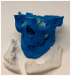
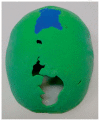
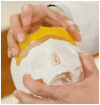
Plastic Surgery restores unique human qualities such as appearance, speech (palate), hands, to improve interaction with others and quality of life. Three-dimensional printing technology can be applied to Plastic Surgery craniomaxillofacial operations to change the bony skeleton of the skull, face, and jaws. Three-dimensional printing for patient-specific applications have four types: Type I contour models, Type II guides, Type III splints, Type IV implants. Plastic Surgery innovation in 3D printing clinical applications are described here and https://www.slucare.edu/newsroom/kmov-science-of-healing-faces-of-childhood.php.
Copyright 2020 by the Missouri State Medical Association.
Figures


References
-
- Jacobs CA, Lin AY. A New Classification of Three-Dimensional Printing Technologies: Systematic Review of Three-Dimensional Printing for Patient-Specific Craniomaxillofacial Surgery. Plast Reconstr Surg. 2017;139(5):1211–1220. - PubMed
-
- Lin AY, Losee JE. Pediatric Plastic Surgery. In: Zitelli BJ, McIntire SC, Nowalk AJ, editors. Zitelli and Davis’ atlas of pediatric physical diagnosis. 6th ed. Philadelphia, PA: Saunders/Elsevier; 2012.
-
- Danelson KA, Gordon ES, David LR, Stitzel JD. Using a three dimensional model of the pediatric skull for pre-operative planning in the treatment of craniosynostosis - biomed 2009. Biomed Sci Instrum. 2009;45:358–363. - PubMed
-
- Engel M, Hoffmann J, Castrillon-Oberndorfer G, Freudlsperger C. The value of three-dimensional printing modelling for surgical correction of orbital hypertelorism. Oral Maxillofac Surg. 2015;19(1):91–95. - PubMed
-
- Hatamleh MM, Cartmill M, Watson J. Management of extensive frontal cranioplasty defects. J Craniofac Surg. 2013;24(6):2018–2022. - PubMed
Publication types
MeSH terms
LinkOut - more resources
Full Text Sources
