

Morphological Analysis of the Human Maxillary Sinus Using Three-Dimensional Printing
- PMID: 32308293
- PMCID: PMC7145240
- DOI: 10.4103/ccd.ccd_548_18
Morphological Analysis of the Human Maxillary Sinus Using Three-Dimensional Printing
Abstract
Background: The maxillary sinus (MS) is described as a pyramid-shaped cavity of the maxilla.
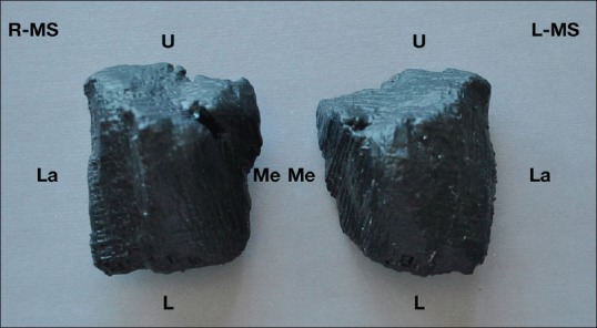
Aim: The aim of this research is to present a strategy for morphological analysis of the MS using three-dimensional (3D) printing acquired through cone-beam computed tomography images.
Material and methods: A cross-sectional exploratory, single-blind study was conducted, including 24 subjects. MSs were reconstructed, and 3D virtual modeling was done bilaterally, obtaining 48 physical models generated on a 3D printer. The statistical analysis used tests of normality and tests using a value of P < 0.05 to establish statistical significance.
Results: The mean of the MS volume was 15.38 cm3 (±6.83 cm3). The minimum volume was 5.4 cm3 and the maximum was 30.8 cm3. In a bilateral comparison of the right and left volume of the same individual, there were no significant differences (P = 0.353). In relation to the morphology of the MSs, the most prevalent was pyramidal with a square base with a prevalence of 66.7%. Related to gender, significant differences were observed only for the left volume (P = 0.009), with the mean volume being significantly greater in the men (19.69 cm3) than in the women (12.28 cm3).
Conclusion: 3D printing of the MS permitted the more precise observation of anatomical features that cannot be seen on a 2D screen. A classification is presented that allows an analysis of sinus morphology, although it is necessary to conduct studies with larger samples to obtain more conclusive results.
Keywords: Maxillary sinus; sinus morphology; three-dimensional printing.
Copyright: © 2020 Contemporary Clinical Dentistry.
Conflict of interest statement
There are no conflicts of interest.
Figures

References
-
- Misch CE, Resnik RR, Misch-Dietsh F. Contemporary Implant Dentistry. 38. Barcelona, España: Elsevier Mosby Inc; 2008. Maxillary sinus anatomy, pathology, and graft surgery; pp. 905–74.
-
- Ryu J, Choi SH, Cha JY, Lee KJ, Hwang CJ. Retrospective study of maxillary sinus dimensions and pneumatization in adult patients with an anterior open bite. Am J Orthod Dentofacial Orthop. 2016;150:796–801. - PubMed
-
- Lawson W, Patel ZM, Lin FY. The development and pathologic processes that influence maxillary sinus pneumatization. Anat Rec (Hoboken) 2008;291:1554–63. - PubMed
-
- Nimigean V, Nimigean VR, Măru N, Sălăvăstru DI, Bădiţă D, Tuculină MJ, et al. The maxillary sinus floor in the oral implantology. Rom J Morphol Embryol. 2008;49:485–9. - PubMed
-
- Jun BC, Song SW, Park CS, Lee DH, Cho KJ, Cho JH. The analysis of maxillary sinus aeration according to aging process; volume assessment by 3-dimensional reconstruction by high-resolutional CT scanning. Otolaryngol Head Neck Surg. 2005;132:429–34. - PubMed
LinkOut - more resources
Full Text Sources