

Virus-Like Particles Presenting the FGF-2 Protein or Identified Antigenic Peptides Promoted Antitumor Immune Responses in Mice
- PMID: 32308382
- PMCID: PMC7146011
- DOI: 10.2147/IJN.S237182
Virus-Like Particles Presenting the FGF-2 Protein or Identified Antigenic Peptides Promoted Antitumor Immune Responses in Mice
Abstract
Background: Fibroblast growth factor (FGF)-2 is overexpressed in various tumor tissues. It affects tumor cell proliferation, invasion and survival, promotes tumor angiogenesis and is tightly involved in the development of systemic and local immunosuppressive tumor mechanisms.
Purpose: This study aimed to develop an effective vaccine against FGF-2 and to investigate the effects of anti-FGF-2 immunization on tumor growth and antitumor immune responses.
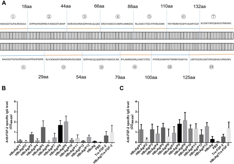
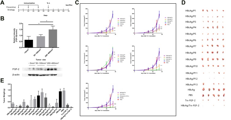
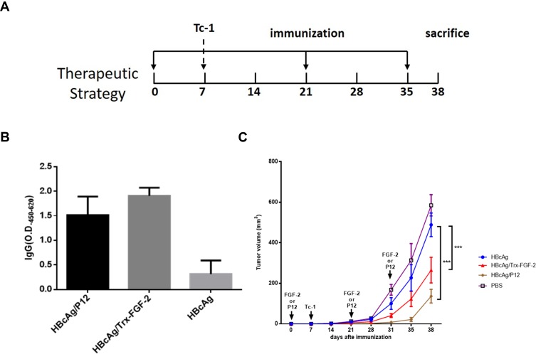
Methods: A set of thirteen synthesized overlapping peptides covering all possible linear B-cell epitopes of murine FGF-2 and a recombinant FGF-2 protein were conjugated to virus-like particles (VLPs) of recombinant hepatitis B core antigen (HBcAg). The VLPs were immunized through a preventive or therapeutic strategy in a TC-1 or 4T1 grafted tumor model.
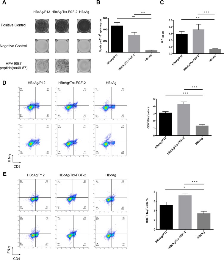
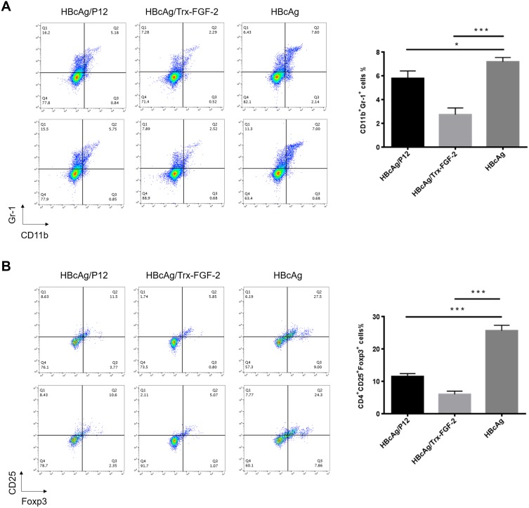
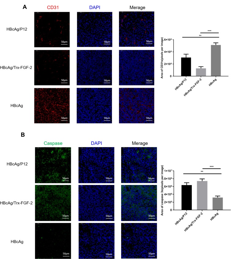
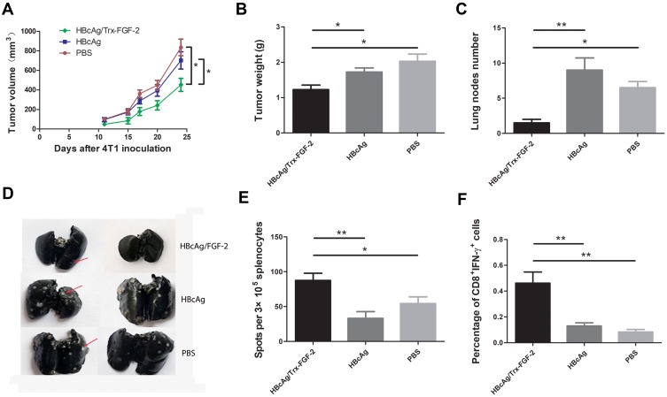
Results: Immunization with FGF-2 peptides or full-length protein-coupled VLPs produced FGF-2-specific antibodies with a high titer. Peptide 12, which is located in the heparin-binding site of FGF-2, or protein-conjugated VLPs presented the most significant effects on the suppression of TC-1 tumor growth. The levels of IFN-γ-expressing splenocytes and serum IFN-γ were significantly elevated; further, the immune effector cells CD8+ IFN-γ+ cytotoxic T lymphocytes (CTLs) and CD4+ IFN-γ+ Th1 cells were significantly increased, whereas the immunosuppressive cells CD4+ CD25+ FOXP3+ Treg cells and Gr-1+ CD11b+ myeloid-derived suppressor cells (MDSCs) were decreased in the immunized mice. In addition, VLP immunization significantly suppressed tumor vascularization and promoted tumor cell apoptosis. In mice bearing 4T1 breast tumor, preventive immunization with FGF-2-conjugated VLPs suppressed tumor growth and lung metastasis, and increased effector cell responses.
Conclusion: Active immunization against FGF-2 is a new possible strategy for tumor immunotherapy.
Keywords: VLP; anticytokine active immunization; fibroblast growth factor-2; immune response; tumor; virus-like particle.
© 2020 Shu et al.
Conflict of interest statement
The authors report no conflicts of interest in this work.
Figures
Similar articles
-
Chimeric HBcAg virus-like particles presenting a HPV 16 E7 epitope significantly suppressed tumor progression through preventive or therapeutic immunization in a TC-1-grafted mouse model.Int J Nanomedicine. 2016 May 27;11:2417-29. doi: 10.2147/IJN.S102467. eCollection 2016. Int J Nanomedicine. 2016. PMID: 27313455 Free PMC article.
-
Recombinant virus-like particles presenting IL-33 successfully modify the tumor microenvironment and facilitate antitumor immunity in a model of breast cancer.Acta Biomater. 2019 Dec;100:316-325. doi: 10.1016/j.actbio.2019.09.024. Epub 2019 Sep 19. Acta Biomater. 2019. PMID: 31542504
-
A therapeutic HPV16 E7 vaccine in combination with active anti-FGF-2 immunization synergistically elicits robust antitumor immunity in mice.Nanomedicine. 2020 Oct;29:102254. doi: 10.1016/j.nano.2020.102254. Epub 2020 Jun 30. Nanomedicine. 2020. PMID: 32615335
-
DNA-mediated immunization to the hepatitis B surface antigen. Activation and entrainment of the immune response.Ann N Y Acad Sci. 1995 Nov 27;772:64-76. doi: 10.1111/j.1749-6632.1995.tb44732.x. Ann N Y Acad Sci. 1995. PMID: 8546414 Review.
-
Virus-like Particle (VLP) Vaccines for Cancer Immunotherapy.Int J Mol Sci. 2023 Aug 19;24(16):12963. doi: 10.3390/ijms241612963. Int J Mol Sci. 2023. PMID: 37629147 Free PMC article. Review.
Cited by
-
Recombinant VLPs empower RBM peptides showing no immunogenicity in native SARS-COV-2 protein to elicit a robust neutralizing antibody response.Nanomedicine. 2022 Apr;41:102527. doi: 10.1016/j.nano.2022.102527. Epub 2022 Jan 30. Nanomedicine. 2022. PMID: 35104670 Free PMC article.
-
Recent Advances and Challenges in Cancer Immunotherapy.Cancers (Basel). 2022 Aug 17;14(16):3972. doi: 10.3390/cancers14163972. Cancers (Basel). 2022. PMID: 36010965 Free PMC article. Review.
-
CD25+FOXP3+CD45RA- regulatory T-cell infiltration as a prognostic biomarker for endometrial carcinoma.BMC Cancer. 2024 Sep 4;24(1):1100. doi: 10.1186/s12885-024-12851-0. BMC Cancer. 2024. PMID: 39232704 Free PMC article.
-
Identification, Characterization and Functional Analysis of Fibroblast Growth Factors in Black Rockfish (Sebastes schlegelii).Int J Mol Sci. 2023 Feb 11;24(4):3626. doi: 10.3390/ijms24043626. Int J Mol Sci. 2023. PMID: 36835037 Free PMC article.
-
P. aeruginosa Mediated Necroptosis in Mouse Tumor Cells Induces Long-Lasting Systemic Antitumor Immunity.Front Oncol. 2021 Feb 12;10:610651. doi: 10.3389/fonc.2020.610651. eCollection 2020. Front Oncol. 2021. PMID: 33643911 Free PMC article.
References
MeSH terms
Substances
LinkOut - more resources
Full Text Sources
Research Materials
