. 2020 Apr 3:11:354.
doi: 10.3389/fpls.2020.00354. eCollection 2020.

SNARE Complexity in Arbuscular Mycorrhizal Symbiosis

Affiliations

SNARE Complexity in Arbuscular Mycorrhizal Symbiosis

Rik Huisman et al. Front Plant Sci. .

Abstract

How cells control the proper delivery of vesicles and their associated cargo to specific plasma membrane (PM) domains upon internal or external cues is a major question in plant cell biology. A widely held hypothesis is that expansion of plant exocytotic machinery components, such as SNARE proteins, has led to a diversification of exocytotic membrane trafficking pathways to function in specific biological processes. A key biological process that involves the creation of a specialized PM domain is the formation of a host-microbe interface (the peri-arbuscular membrane) in the symbiosis with arbuscular mycorrhizal fungi. We have previously shown that the ability to intracellularly host AM fungi correlates with the evolutionary expansion of both v- (VAMP721d/e) and t-SNARE (SYP132α) proteins, that are essential for arbuscule formation in Medicago truncatula. Here we studied to what extent the symbiotic SNAREs are different from their non-symbiotic family members and whether symbiotic SNAREs define a distinct symbiotic membrane trafficking pathway. We show that all tested SYP1 family proteins, and most of the non-symbiotic VAMP72 members, are able to complement the defect in arbuscule formation upon knock-down/-out of their symbiotic counterparts when expressed at sufficient levels. This functional redundancy is in line with the ability of all tested v- and t-SNARE combinations to form SNARE complexes. Interestingly, the symbiotic t-SNARE SYP132α appeared to occur less in complex with v-SNAREs compared to the non-symbiotic syntaxins in arbuscule-containing cells. This correlated with a preferential localization of SYP132α to functional branches of partially collapsing arbuscules, while non-symbiotic syntaxins accumulate at the degrading parts. Overexpression of VAMP721e caused a shift in SYP132α localization toward the degrading parts, suggesting an influence on its endocytic turn-over. These data indicate that the symbiotic SNAREs do not selectively interact to define a symbiotic vesicle trafficking pathway, but that symbiotic SNARE complexes are more rapidly disassembled resulting in a preferential localization of SYP132α at functional arbuscule branches.

Keywords: Medicago; SNARE; VAMP; arbuscular mycorrhiza; exocytosis; membrane; symbiosis; syntaxin.


Figures

FIGURE 1
FIGURE 1
The SNARE repertoire of arbuscule containing cells. (A) Schematic phylogenetic trees showing the evolution of the exocytosis related SNARE families SYP1 and VAMP72. The trees are showing orthogroups instead of individual genes, indicated by the use of roman numbers. Orthogroups were named following Sanderfoot (2007). Both schematic trees are based on the actual phylogenetic trees shown in Supplementary Figures S1, S2. (B) Digital droplet PCR on cDNA from arbuscule containing cells. Only SYP1 and VAMP72 genes that are expressed in arbuscule-containing cells are shown. The copy in each cDNA was normalized against the average expression of all tested genes. Error bars represent standard deviation of three biological replicates.
FIGURE 2
FIGURE 2
Intracellular localization of syntaxins (A–D) and VAMPs (E–H) in arbuscule containing cells. Different SNAREs fused to GFP were expressed from the arbuscule specific Medicago PT4 promoter, combined with dsRed that marks the cytoplasm and nucleus (n), as well as occasional accumulation in the vacuoles (v). white arrowheads indicate the PAM, a green arrow indicates the tonoplast. Scalebars are 10 μm.
FIGURE 3
FIGURE 3
Co-immunoprecipitation analysis of SNARE interactions. Western blot showing GFP-labeled syntaxins or negative control PT1, and 3HA-labeled VAMPs in extracts of mycorrhized Medicago roots before (input) and after immunoprecipitation (IP) using anti-GFP coated beads. The fraction (% HA retrieval) of HA-labeled VAMPs that co-purified with the GFP-labeled bait proteins was quantified as the intensity of the bands in the IP lane, divided by the intensity of the corresponding band in the input lane, and corrected for the volume difference of the two fractions.
FIGURE 4
FIGURE 4
RNAi complementation analysis. Projection of confocal image stacks showing AM fungi in Medicago roots, stained with wheat germ agglutinin conjugated to alexa488 n = 3–4 biological replicates. (A) Empty vector control, (B) RNAi of SYP132α, (C–F) complementation of SYP132 RNAi with other syntaxins, (G) RNAi of VAMP721d and -e, and (H–K) complementation of VAMP721d/e RNAi with other VAMPs.
FIGURE 5
FIGURE 5
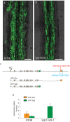
(A,B) Mycorrhized wild-type (A) and syp132α-1 roots stained with WGA-Alexa488. Scale bars are 100 μm. (C) Schematic representation of the SYP132 exons in the genome of Medicago, the different spliced transcripts, and the region deleted in the syp132α-1 mutant. (D) Digital droplet PCR measurements of the transcript levels of SYP132α and β in the arbuscule-containing cells of wild-type plants and syp132α-1 plants. Error bars represent standard deviation of 4 (R108) or 3 (syp132α-1) biological replicates.
FIGURE 6
FIGURE 6
(A) Colonization (M%) and arbuscule abundance (A%) in wild-type and syp132α-1 roots, 6 weeks after inoculation with R. irregularis. Parameters are scored according to Trouvelot et al. (1986). Error bars represent the standard deviation of five biological replicates. (B) qRT-PCR on cDNA isolated from mycorrhized roots of wild-type and syp132α-1 showing the expression of marker gene PT4. Error bars represent the standard deviation of three biological replicates. (C) Arbuscule length distribution measured in wild-type and syp132α-1 roots infected with R. irregularis. Around 1000 arbuscules per root were measured. Error bars represent standard deviation of four different roots. (D) Confocal laser scanning microscopy images overlaid with DIC images showing expression of nuclear localized dsRed-E5 (Timer) driven by the PT4 promoter in arbuscular cells of wild-type plants. The numbers in the upper-right corner indicate the ratio between red and green fluorescence. (E) Age distribution of arbuscules in wild-type and syp132α-1 roots. The age of ∼100 arbuscules was measured per root. Collapsed arbuscules were not measured. Error bars represent standard deviation of four individual roots. (A–C,E) No significant differences between wild-type and syp132α-1 were found (Student’s t-test p ≤ 0.05).
FIGURE 7
FIGURE 7
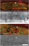
Confocal laser scanning microscopy images and accompanying bright-field images showing the localization of SYP132α fused to GFP with (A) and without (B) co-expression of triple HA-tag labeled VAMP721e, both driven by the PT4 promoter. Collapsed branches are marked by white arrowheads, functional branches are marked by blue arrowheads. The red signal shows autofluorescence of collapsed arbuscules and the nuclei and cytoplasm that are labeled with dsRed driven by the Arabidopsis ubiquitin 3 promoter. Scale bars are 10 μm.

References

    1. An Q., Hückelhoven R., Kogel K. H., van Bel A. J. E. (2006). Multivesicular bodies participate in a cell wall-associated defence response in barley leaves attacked by the pathogenic powdery mildew fungus. Cell. Microbiol. 8 1009–1019. 10.1111/j.1462-5822.2006.00683.x - DOI - PubMed
    1. Ângelo Silva P., Ul-Rehman R., Rato C., Di Sansebastiano G.-P., Malhó R. (2010). Asymmetric localization of Arabidopsis SYP124 syntaxin at the pollen tube apical and sub-apical zones is involved in tip growth. BMC Plant Biol. 10:179. 10.1186/1471-2229-10-179 - DOI - PMC - PubMed
    1. Assaad F. F., Qiu J. L., Youngs H., Ehrhardt D., Zimmerli L., Kalde M., et al. (2004). The PEN1 syntaxin defines a novel cellular compartment upon fungal attack and is required for the timely assembly of papillae. Mol. Biol. Cell 15 5118–5129. 10.1091/mbc.e04-02-0140 - DOI - PMC - PubMed
    1. Bravo A., York T., Pumplin N., Mueller L. A., Harrison M. J. (2016). Genes conserved for arbuscular mycorrhizal symbiosis identified through phylogenomics. Nat. Plants 2:15208. 10.1038/nplants.2015.208 - DOI - PubMed
    1. Chabaud M., De Carvalho-Niebel F., Barker D. G. (2003). Efficient transformation of Medicago truncatula cv. Jemalong using the hypervirulent Agrobacterium tumefaciens strain AGL1. Plant Cell Rep. 22 46–51. 10.1007/s00299-003-0649-y - DOI - PubMed

LinkOut - more resources