Review
. 2020 Apr 6;15(2):187-194.
doi: 10.18502/jovr.v15i2.6736. eCollection 2020 Apr-Jun.

Torpedo Retinopathy

Affiliations
Review

Torpedo Retinopathy

Ramesh Venkatesh et al. J Ophthalmic Vis Res. .

Abstract

Purpose: Torpedo lesions in the retina are rare. This study aimed to investigate torpedo-shaped lesions in the retina in an adult population and to determine the spectrum and features of the disease.

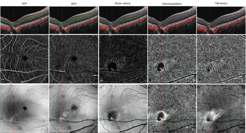
Methods: The review of a database for clinical diagnosis identified nine patients who were diagnosed with torpedo-shaped lesions in the retina between June 2017 and February 2019. Fundus photography and optical coherence tomography (OCT) imaging were used to analyze the cases. Multicolor imaging was also performed.

Results: Nine patients with torpedo-shaped lesions in the fundus were identified. Fundus images revealed that the lesion involved the macula in six eyes; in the remaining three eyes, the lesion was present outside the macula. OCT identified six patients with type 1 torpedo lesions, one with type 2, and two with type 3. On multicolor imaging, the lesion was visualized as a region of increased reflectance in blue, green, and infrared light in all eyes, with notably increased infrared reflectance in eyes with focal choroidal excavation. Choroidal neovascular membrane was evident in one patient on OCT angiography.

Conclusion: Torpedo lesions in the retina can occur away from the macula and exhibit features similar to those of torpedo maculopathy. As such, the authors propose a change in the nomenclature for torpedo lesions in the retina from "torpedo maculopathy" to "torpedo retinopathy."

Keywords: Maculopathy; Retinopathy; Torpedo; Imaging.


Conflict of interest statement

There are no conflicts of interest.

Figures

Figure 1
Figure 1
Fundus and OCT imaging of torpedo lesions (patients 1–9). The torpedo lesion in each panel is marked on the fundus image with white arrow. In panels, C, F, G and I, the torpedo lesion is located adjacent to the fovea. In panels B and E, the lesion is located adjacent to the macula but away from the fovea. In panel A, the lesion is just within the retinal arcade, and in panels D and H, the lesion is outside the retinal arcade. In every case, the OCT scan passing through the torpedo lesion is depicted.
Figure 2
Figure 2
OCT angiography findings of patient 6 showing the torpedo lesion with focal choroidal excavation with presence of choroidal neovascular membrane. Columns 1–5 show the automated segmentation scans through the superficial capillary plexus, deep capillary plexus, outer retina, choriocapillaris, and manual segmentation across the torpedo lesion with focal choroidal excavation, respectively. Note the decreased flow areas and vessel density in the area of torpedo lesion. A choroidal neovascular membrane can be identified as well.
Figure 3
Figure 3
Adaptive optics imaging of the right eye in patient 3 with torpedo lesion. Raw images on adaptive optics imaging showing the exact site of cone density and spacing analysis (bold white arrow). Adaptive optics imaging showing raw images, cone density, and cone spacing of right eye at 2° from the foveal center in all four quadrants.

References

    1. Villegas Victor M., Schwartz Stephen G., Flynn Harry W., Capó Hilda, Berrocal Audina M., Murray Timothy G., Harbour J. William. Distinguishing Torpedo Maculopathy From Similar Lesions of the Posterior Segment. Ophthalmic Surgery, Lasers and Imaging Retina. 2014;45(3):222–226. doi: 10.3928/23258160-20140410-01. - DOI - PubMed
    1. Gass J. Donald M. Solitary Hypopigmented Nevus of the Retinal Pigment Epithelium in the Macula. Archives of Ophthalmology. 1992;110(10):1358. doi: 10.1001/archopht.1992.01080220020005. - DOI - PubMed
    1. Golchet P. R, Jampol L. M, Mathura J. R, Daily M. J. Torpedo maculopathy. British Journal of Ophthalmology. 2009;94(3):302–306. doi: 10.1136/bjo.2009.162669. - DOI - PubMed
    1. Traboulsi Elias I. Pigmented and depigmented lesions of the ocular fundus. Current Opinion in Ophthalmology. 2012;23(5):337–343. doi: 10.1097/icu.0b013e32835622b0. - DOI - PubMed
    1. Traboulsi E. Ocular Manifestations of Familial Adenomatous Polyposis (Gardner Syndrome) Ophthalmology Clinics of North America. 2005;18(1):163–166. doi: 10.1016/j.ohc.2004.08.003. - DOI - PubMed

LinkOut - more resources