Proliferating Infantile Hemangioma Tissues and Primary Cell Lines Express Markers Associated with Endothelial-to-Mesenchymal Transition
- PMID: 32309069
- PMCID: PMC7159972
- DOI: 10.1097/GOX.0000000000002598
Proliferating Infantile Hemangioma Tissues and Primary Cell Lines Express Markers Associated with Endothelial-to-Mesenchymal Transition
Abstract
Background: We have previously shown that the endothelium of the microvessels of infantile hemangioma (IH) exhibits a hemogenic endothelium phenotype and proposed its potential to give rise to mesenchymal stem cells, similar to the development of hematopoietic cells. This endothelial-to-mesenchymal transition (Endo-MT) process involves the acquisition of a migratory phenotype by the endothelial cells, similar to epithelial-to-mesenchymal transition that occurs during neural crest development. We hypothesized that proliferating IH expresses Endo-MT-associated proteins and investigated their expression at the mRNA, protein, and functional levels.
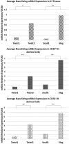
Methods: Immunohistochemical staining of paraffin-embedded sections of proliferating IH samples from 10 patients was undertaken to investigate the expression of the Endo-MT proteins Twist1, Twist2, Snail1, and Slug. Transcriptional analysis was performed for the same markers on proliferating IH tissues and CD34+ and CD34- cells from proliferating IH-derived primary cell lines. Adipogenic and osteogenic differentiation plasticity was determined on the CD34-sorted fractions.
Results: The endothelium of the microvessels and the cells within the interstitium of proliferating IH tissues expressed Twist1, Twist2, and Slug proteins. Twist1 was also expressed on the pericyte layer of the microvessels, whereas Snail1 was not expressed. Both CD34+ and CD34- populations from the IH-derived primary cell lines underwent adipogenic and osteogenic differentiation.
Conclusions: The expression of Endo-MT-associated proteins Twist1, Twist2, and Slug by both the endothelium of the microvessels and cells within the interstitium, and Twist1 on the pericyte layer of the microvessels of proliferating IH, suggest the presence of a process similar to Endo-MT. This may enable a tightly controlled primitive endothelium of proliferating IH to acquire a migratory mesenchymal phenotype with the ability to migrate away, providing a plausible explanation for the development of a fibrofatty residuum observed during involution of IH.
Copyright © 2020 The Authors. Published by Wolters Kluwer Health, Inc. on behalf of The American Society of Plastic Surgeons.
Figures


Similar articles
-
Infantile haemangioma expresses embryonic stem cell markers.J Clin Pathol. 2012 May;65(5):394-8. doi: 10.1136/jclinpath-2011-200462. Epub 2012 Mar 23. J Clin Pathol. 2012. PMID: 22447921
-
Expression of (pro)renin receptor and its effect on endothelial cell proliferation in infantile hemangioma.Pediatr Res. 2019 Aug;86(2):202-207. doi: 10.1038/s41390-019-0430-8. Epub 2019 May 15. Pediatr Res. 2019. PMID: 31091531
-
Biology of infantile hemangioma.Front Surg. 2014 Sep 25;1:38. doi: 10.3389/fsurg.2014.00038. eCollection 2014. Front Surg. 2014. PMID: 25593962 Free PMC article. Review.
-
R(+) Propranolol decreases lipid accumulation in hemangioma-derived stem cells.bioRxiv [Preprint]. 2024 Jul 2:2024.07.01.601621. doi: 10.1101/2024.07.01.601621. bioRxiv. 2024. Update in: Br J Dermatol. 2025 Mar 18;192(4):757-759. doi: 10.1093/bjd/ljae452. PMID: 39005472 Free PMC article. Updated. Preprint.
-
Cardiovascular drugs in the treatment of infantile hemangioma.World J Cardiol. 2016 Jan 26;8(1):74-80. doi: 10.4330/wjc.v8.i1.74. World J Cardiol. 2016. PMID: 26839658 Free PMC article. Review.
Cited by
-
Pathogenesis of Cardiac Valvular Hemangiomas: A Case Report and Literature Review.Int J Mol Sci. 2025 Jul 23;26(15):7114. doi: 10.3390/ijms26157114. Int J Mol Sci. 2025. PMID: 40806246 Free PMC article. Review.
References
-
- Amrock SM, Weitzman M. Diverging racial trends in neonatal infantile hemangioma diagnoses, 1979-2006. Pediatr Dermatol. 2013;30:493–494. - PubMed
-
- Hoornweg MJ, Smeulders MJ, Ubbink DT, et al. The prevalence and risk factors of infantile haemangiomas: a case-control study in the Dutch population. Paediatr Perinat Epidemiol. 2012;26:156–162. - PubMed
-
- Tan ST, Itinteang T, Leadbitter P. Low-dose propranolol for infantile haemangioma. J Plast Reconstr Aesthet Surg. 2011;64:292–299. - PubMed
-
- Mulliken JB, Glowacki J. Hemangiomas and vascular malformations in infants and children: a classification based on endothelial characteristics. Plast Reconstr Surg. 1982;69:412–422. - PubMed
-
- Itinteang T, Tan ST, Jia J, et al. Mast cells in infantile haemangioma possess a primitive myeloid phenotype. J Clin Pathol. 2013;66:597–600. - PubMed
LinkOut - more resources
Full Text Sources
Research Materials
