

Amniotic membrane transplantation for managing dry eye and neurotrophic keratitis
- PMID: 32309119
- PMCID: PMC7158925
- DOI: 10.4103/tjo.tjo_5_20
Amniotic membrane transplantation for managing dry eye and neurotrophic keratitis
Abstract
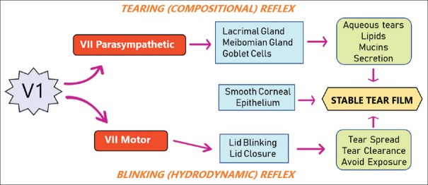
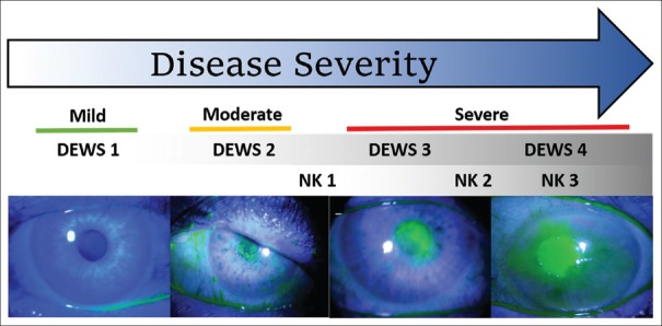
Neurotrophic keratitis (NK), a degenerative disease caused by damage to the trigeminal nerve, abolishes both tearing and blinking reflexes, thus causing the most severe forms of dry eye disease (DED). Conversely, the increasing severity of DED also leads to progressive loss of corneal nerve density, potentially resulting in NK. Both diseases manifest the same spectrum of corneal pathologies including inflammation and corneal epithelial keratitis, which can progress into vision-threatening epithelial defect and stromal ulceration. This review summarizes the current literature regarding outcomes following sutured and sutureless cryopreserved amniotic membrane (AM) in treating DED as well as epithelial defects and corneal ulcers due to underlying NK. These studies collectively support the safety and effectiveness of cryopreserved AM in restoring corneal epithelial health, improving visual acuity in eyes with NK and DED, and alleviating symptomatic DED. Future randomized controlled trials are warranted to validate the above findings and determine whether such clinical efficacy lies in promoting corneal nerve regeneration.
Keywords: Amniotic membrane; corneal ulcer; dry eye; epithelial defect; neurotrophic keratitis.
Copyright: © 2020 Taiwan J Ophthalmol.
Conflict of interest statement
OM, ST, and ST are employees of TissueTech. Dr Tseng has obtained a patent for the method of preparation and clinical uses of amniotic membrane and has licensed the rights to TissueTech, Inc, which procures and processes, and to Bio-Tissue, Inc, which is a subsidiary of TissueTech, Inc, to distribute cryopreserved amniotic membrane for clinical and research uses.
Figures
References
-
- Okada Y, Sumioka T, Ichikawa K, Sano H, Nambu A, Kobayashi K, et al. Sensory nerve supports epithelial stem cell function in healing of corneal epithelium in mice: The role of trigeminal nerve transient receptor potential vanilloid 4. Lab Invest. 2019;99:210–30. - PubMed
-
- Yang L, Di G, Qi X, Qu M, Wang Y, Duan H, et al. SubstancePpromotes diabetic corneal epithelial wound healing through molecular mechanisms mediated via the neurokinin-1 receptor. Diabetes. 2014;63:4262–74. - PubMed
-
- Mikulec AA, Tanelian DL. CGRP increases the rate of corneal re-epithelialization in an in vitro whole mount preparation. J Ocul Pharmacol Ther. 1996;12:417–23. - PubMed
Publication types
LinkOut - more resources
Full Text Sources