

Quantitative analysis by reversed-phase high-performance liquid chromatography and retinal neuroprotection after topical administration of moxonidine
- PMID: 32309174
- PMCID: PMC7154207
- DOI: 10.18240/ijo.2020.03.04
Quantitative analysis by reversed-phase high-performance liquid chromatography and retinal neuroprotection after topical administration of moxonidine
Abstract
Aim: To determine moxonidine in aqueous humor and iris-ciliary body by reversed-phase high performance liquid chromatography (RP-HPLC), and to evaluate the retinal neuroprotective effect after topical administration with moxonidine in a high intraocular pressure (IOP) model.
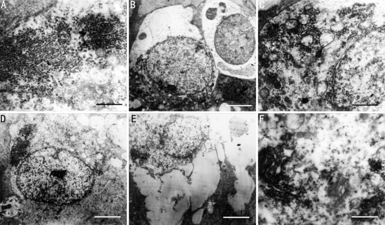
Methods: The eyes of albino rabbits were administered topically and ipsilaterally with 0.2% moxonidine. A RP-HPLC method was employed for the identification and quantification of moxonidine between 2 and 480min, which presented in the aqueous humor and iris-ciliary body. Flash electroretinography (F-ERG) amplitude and superoxide dismutase (SOD) level were measured between day 1 and day 15 after topical administration with moxonidine in a rabbit model of high IOP. Histological and ultrastructural observation underwent to analyze the changes of retinal morphology, the inner retinal layers (IRL) thickness, and retinal ganglion cell (RGC) counting.
Results: Moxonidine was detectable between 2 and 480min after administration, and the peak concentration developed both in the two tissues at 30min, 0.51 µg/mL in aqueous humor and 1.03 µg/g in iris-ciliary body. In comparison to control, F-ERG b-wave amplitude in moxonidine eyes were significantly differences between day 3 and day 15 (P<0.01) in the high IOP model; SOD levels were significantly higher at all time-points (P<0.01) with a maximum level of 20.29 U/mgprot at day 15; and RGCs were significantly higher (P<0.05).
Conclusion: Moxonidine is a viable neuroprotective agent with application to high IOP model. All layers of retina, including RGC layer, retinal nerve fiber layer and INL, are more preserved after moxonidine administration. SOD plays a neuroprotective role in ocular hypertension-mediated RGC death.
Keywords: moxonidine; neuroprotection; retinal ganglion cell; reversed-phase high-performance liquid chromatography; superoxide dismutase.
International Journal of Ophthalmology Press.
Figures


References
-
- Cobos-Puc LE, Aguayo-Morales H, Silva-Belmares Y, González-Zavala MA, Centurión D. Α2A-adrenoceptors, but not nitric oxide, mediate the peripheral cardiac sympatho-inhibition of moxonidine. Eur J Pharmacol. 2016;782:35–43. - PubMed
-
- Soldatov VO, Shmykova EA, Pershina MA, Ksenofontov AO, Zamitsky YM, Kulikov AL, Peresypkina AA, Dovgan AP, Belousova YV. Imidazoline receptors agonists: possible mechanisms of endothelioprotection. Res Results Pharmacol. 2018;4(2):11–19.
-
- Zhang LL, Ding L, Zhang F, Gao R, Chen Q, Li YH, Kang YM, Zhu GQ. Salusin-β in rostral ventrolateral medulla increases sympathetic outflow and blood pressure via superoxide anions in hypertensive rats. J Hypertens. 2014;32(5):1059–1067. discussion 1067. - PubMed
-
- Yang JG, Sun NX, Xiong QC, Yang R. Effect of moxonidine on the uveoscleral outflow: role of alpha2-adrenoceptors or I1 imidazoline receptors. Curr Eye Res. 2009;34(4):287–296. - PubMed
-
- Barygina V, Becatti M, Lotti T, Moretti S, Taddei N, Fiorillo C. ROS-challenged keratinocytes as a new model for oxidative stress-mediated skin diseases. J Cell Biochem. 2019;120(1):28–36. - PubMed
LinkOut - more resources
Full Text Sources