. 2020 Mar;8(5):193.
doi: 10.21037/atm.2020.01.63.

Pathological myopia-induced antioxidative proteins in the vitreous humor

Affiliations

Pathological myopia-induced antioxidative proteins in the vitreous humor

Qiaoling Wei et al. Ann Transl Med. 2020 Mar.

Abstract

Background: This study aimed to investigate differentially expressed proteins in the vitreous humor (VH) of pathological myopia (PM) and normal eyes.

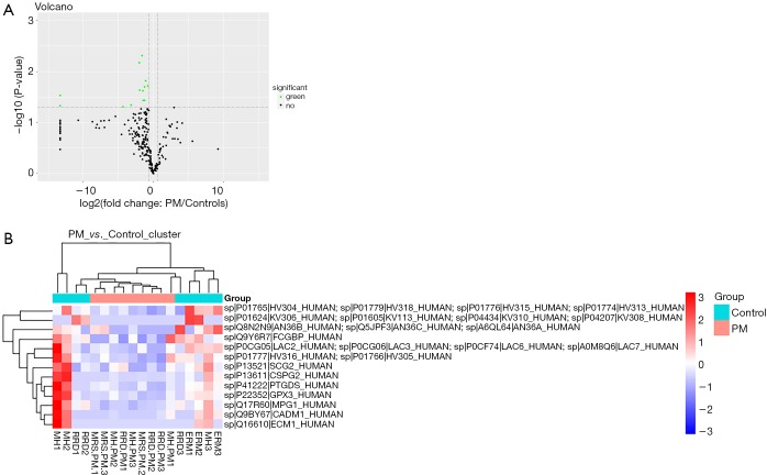
Methods: VH samples were collected from patients undergoing surgical treatment for rhegmatogenous retinal detachment (RRD), idiopathic epiretinal membrane (ERM), myopic retinoschisis (MRS) or macular hole (MH). A label-free quantitative proteomic analysis was performed to detect the differentially expressed proteins, and expression of three differentially expressed proteins was confirmed by ELISA.

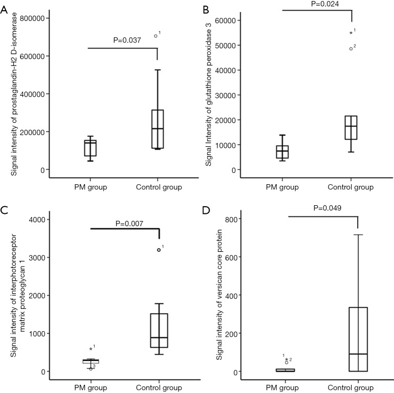
Results: In PM patients (MH-PM, MRS-PM and RRD-PM), the expression of prostaglandin-H2 D-isomerase (PGDS) and glutathione peroxidase 3 (GPX3) was significantly lower than in controls (MH, ERM, and RRD). The versican core protein expression decreased in the PM group. The vitreous concentrations of PGDS and GPX3 in patients with axial length (AL) of 26.5-29.0 mm were higher than in patients with AL >29.0 mm or AL <26.5 mm. NRF-2 expression was the lowest in patients with AL >29.0 mm.

Conclusions: Our study provides new evidence on the molecular changes in the VH of PM patients, and these molecules have the potential to become new targets for the therapy of PM.

Keywords: Pathological myopia (PM); antioxidative protein; prostaglandin-H2 d-isomerase (PGDS); retinal pigment epithelium (RPE).


Conflict of interest statement

Conflicts of Interest: The authors have no conflicts of interest to declare.

Figures

Figure 1
Figure 1
Comparison of PM patients and controls. (A) Signal intensity of prostaglandin-H2 d-isomerase; (B) signal intensity of glutathione peroxidase 3; (C) signal intensity of interphotoreceptor matrix proteoglycan 1; (D) signal intensity of versican core protein; Signal intensities were determined by label-free quantitative proteomics analysis. Student’s t-test was used to compare the signal intensities of differentially expressed proteins between 2 groups. PM, pathological myopia.
Figure 2
Figure 2
Differentially expressed proteins in the VH of PM patients and controls (label free quantitative proteomics analysis). (A) Volcano plot represents the differentially expressed protein. The −log10 (P value) is plotted against the difference of the means of two groups (PM and controls). Dots above the non-axial horizontal line are significantly differentially expressed proteins. Thirteen proteins were found to be significantly (P<0.002) downregulated in the PM samples (green plots), while none was upregulated (red plots), and the proteins with unchanged expression were marked with dark dots. (B) Hierarchical clustering analysis of 13 differentially expressed proteins between PM patients and controls. Protein names are listed on the right, group names are indicated at the top while the subgroups are listed at the bottom. The color key on the left of the heatmap indicates the expression level (red = up-regulation, blue = downregulation). VH, vitreous humor; PM, pathological myopia; ERM, epiretinal membrane; MH, macular hole; RRD, rhegmatogenous retinal detachment; MRS, myopic retinoschisis.

References

    1. Foster PJ, Jiang Y. Epidemiology of myopia. Eye (Lond) 2014;28:202-8. 10.1038/eye.2013.280 - DOI - PMC - PubMed
    1. Wong TY, Ferreira A, Hughes R, et al. Epidemiology and disease burden of pathologic myopia and myopic choroidal neovascularization: an evidence-based systematic review. Am J Ophthalmol 2014;157:9-25.e12. 10.1016/j.ajo.2013.08.010 - DOI - PubMed
    1. Chan NS, Teo K, Cheung CM. Epidemiology and Diagnosis of Myopic Choroidal Neovascularization in Asia. Eye Contact Lens 2016;42:48-55. 10.1097/ICL.0000000000000201 - DOI - PubMed
    1. Koh V, Yang A, Saw SM, et al. Differences in prevalence of refractive errors in young Asian males in Singapore between 1996-1997 and 2009-2010. Ophthalmic Epidemiol 2014;21:247-55. 10.3109/09286586.2014.928824 - DOI - PubMed
    1. Morgan IG, Ohno-Matsui K, Saw SM. Myopia. Lancet 2012;379:1739-48. 10.1016/S0140-6736(12)60272-4 - DOI - PubMed