Review
. 2017 Oct 16;5(4):e79.
doi: 10.15190/d.2017.9.

Functional Amyloids and their Possible Influence on Alzheimer Disease

Affiliations
Review

Functional Amyloids and their Possible Influence on Alzheimer Disease

Angus Lau et al. Discoveries (Craiova). .

Abstract

Amyloids play critical roles in human diseases but have increasingly been recognized to also exist naturally. Shared physicochemical characteristics of amyloids and of their smaller oligomeric building blocks offer the prospect of molecular interactions and crosstalk amongst these assemblies, including the propensity to mutually influence aggregation. A case in point might be the recent discovery of an interaction between the amyloid β peptide (Aβ) and somatostatin (SST). Whereas Aβ is best known for its role in Alzheimer disease (AD) as the main constituent of amyloid plaques, SST is intermittently stored in amyloid-form in dense core granules before its regulated release into the synaptic cleft. This review was written to introduce to readers a large body of literature that surrounds these two peptides. After introducing general concepts and recent progress related to our understanding of amyloids and their aggregation, the review focuses separately on the biogenesis and interactions of Aβ and SST, before attempting to assess the likelihood of encounters of the two peptides in the brain, and summarizing key observations linking SST to the pathobiology of AD. While the review focuses on Aβ and SST, it is to be anticipated that crosstalk amongst functional and disease-associated amyloids will emerge as a general theme with much broader significance in the etiology of dementias and other amyloidosis.

Keywords: Alzheimer disease; Aβ; amyloid; interaction; oligomeric; somatostatin.


Conflict of interest statement

Conflict of interests: A provisional US patent (application number 62/451-309) titled ‘Oligomeric Abeta-Binding Polypeptides) that lists HW and GS as inventors was filed on January 27, 2017, by the University of Toronto.

Figures

Figure 1
Figure 1. Fibrillogenesis and the conditions that promote specific Aß42 assemblies
A. The general flow of fibrillogenesis begins with monomers, which assemble into oligomers, and eventual turn into amyloid protofibrils and fibrils. In addition to the canonical build-up indicated by arrows pointing right, oligomers, and even fibrils, may shed monomers (or oligomeric building blocks) depicted by reverse arrows. B. Flow-chart summaring major Aβ assembly pathways reported in the literature. Key features of the respective protocols are outlined in the flow chart. Roman numerals i.-iii. associate a given conformer with three-dimensional representations depicted in panel A. 12-mer globulomers and ADDLs are associated with the identifier ‘iii’ as they’re hypothesized to form through stacking of planar 6-mers. 5-mer and 10-mer assemblies were also identified with ‘iii’ as they’re thought to form similar structures, albeit with one less protomer. High temperature (T°) denotes a temperature of 37 °C, low T° denotes a range of 4-8 °C. The abbreviation ‘mo.’ denotes months and DPC micelles/bicelles refer to dodecylphosphocholine micelles/bicelles. Red and black (or grey) colors designate toxic and innocuous molecular species, respectively.
Figure 2
Figure 2. Strains, cross-seeding and co-aggregation. Cartoon depicting three related, yet distinct, concepts relevant to protein aggregation phenomena
A. The same protein can give rise to distinct ‘strains’ of protein aggregates, if the arrangement of its monomers and the conformations adopted by the monomers within the aggregates are different. B. The term ‘cross-seeding’ designates a phenomenon whereby small aggregates composed of a given protein can seed the aggregation of another protein, thereby often influencing the kinetics and quaternary structure of aggregates forming. C. When two or more proteins ‘co-aggregate’ their monomers influence each other’s aggregation. Two separate scenarios can be further distinguished: the two different proteins can polymerize together and form mixed molecular species hybrid structures, or they can interact and influence each other’s aggregation, but polymerize separately to form single species structures.
Figure 3
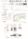
Figure 3. Discovery and validation of SST-Aβ interaction
A. Sequence alignment of preprocortistatin and preprosomatostatin. The signal sequence and the boundaries of the bioactive cortistatin and somatostatin peptides are indicated by horizontal bars. Identical residues are highlighted by black background shading, and peptide sequences observed by mass spectrometry are shown in colored fonts. B. Expanded view of MS3 spectrum derived from ‘NFFWK’ parent spectrum (shown to the right) in interactome study based on oAβ1-42-biotin baits and mAβ1-42-biotin negative controls. In this view, the relative intensities of tandem mass tag (TMT) signature ions reflect the relative abundances of the ‘NFFWK’ peptide in side-by-side generated affinity purification eluate fractions, indicating preferential binding of SST to pre-aggregated oAβ1-42. C. Example tandem MS spectrum supporting the identification of the peptide with amino acid sequence ‘NFFWK’. Fragment masses attributed to B- and Y- ion series are shown in red and blue colors, respectively. D. Workflow of ThT-based aggregation assay. E. SST14 delays Aβ1-42 aggregation in ThT fluorescence assay in a SST14 concentration dependent manner. F. Immunoblot analyses with an antibody directed against an N-terminal Aβ epitope (6E10) reveal that CST17 (or SST14) co-assemble with Aβ1-42 into oligomers of 50-55 kDa that withstand boiling (lanes 2 and 3) but partially disintegrate in the presence of SDS. Note bands of 5-6 kDa, consistent with the existence of SDS-resistant heterodimeric complexes of mAβ1-42 and SST14 (or CST17), and the well-defined oligomeric bands of 50 and 55 kDa (lanes 6 and 7) that were observed in samples derived from the co-incubation of SST14 (or CST17) with Aβ1-42, but not Aβ1-40 (lanes 6, 7, 14, 15). Note also that signals interpreted to represent trimeric Aβ1-42, but not dimeric Aβ1-42, can be seen to migrate slower in the presence of SST14 (or CST17) but not the negative control peptide AVP (compare lanes 9 and 12 with lanes 10 and 11). Finally, intensity levels of homodimeric Aβ1-42 bands are reduced in the presence of SST14 (or CST17) (compare lanes 13 and 16 with lanes 14 and 15). Black arrowhead labeled with ‘m’, ‘d’, and ‘t’ designate bands interpreted to consist of monomeric, dimeric and trimeric Aβ1-42. Green and red arrowheads were used to label bands interpreted to represent SDS-stable heteromeric building blocks consisting of SST14 (or CST17) bound to monomeric and trimeric Aβ1-42, respectively. Elements from this image were adapted from, licensed under CC BY 4.0.
Figure 4
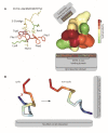
Figure 4. Interactions of cyclic and non-cyclic somatostatin
A. The natural state of somatostatin is cyclic (SST14 and SST28), formed by the presence of a disulfide bridge between cysteine 3 and 14. Cyclic somatostatin is able to dock to sst1-5 receptors with high affinity through a binding epitope spanning residues 7 through 10, which represents the core region responsible for its binding to Aβ. B. Cyclic somatostatin can also form amyloids through self-aggregation. Cysteamine reduces the disulfide bridge, leading to a transition to non-cyclic somatostatin, which has a higher propensity to self-aggregate. The amyloidogenic binding domain in non-cyclic somatostatin is between residues 3 through 14. Adapted from the somatostatin structure in the PDB database.
Figure 5
Figure 5. Widespread distribution of AβPP/Aβ, SST and CST across the human brain
Schematic summarizing key brain areas reported to express AβPP/Aβ, SST and CST or sst1-5 receptors. Bolded labels indicate areas of the brain known to express at least two of the proteins/peptides of interest.

References

    1. The amyloid state and its association with protein misfolding diseases. Knowles Tuomas P. J., Vendruscolo Michele, Dobson Christopher M. Nature Reviews Molecular Cell Biology. 2014;15(6):384-396. - PubMed
    1. Common Structure of Soluble Amyloid Oligomers Implies Common Mechanism of Pathogenesis. Kayed R. Science. 2003;300(5618):486-489. - PubMed
    1. Prefibrillar Amyloid Protein Aggregates Share Common Features of Cytotoxicity. Bucciantini Monica, Calloni Giulia, Chiti Fabrizio, Formigli Lucia, Nosi Daniele, Dobson Christopher M., Stefani Massimo. Journal of Biological Chemistry. 2004;279(30):31374-31382. - PubMed
    1. Alzheimer's disease: Initial report of the purification and characterization of a novel cerebrovascular amyloid protein. Glenner George G., Wong Caine W. Biochemical and Biophysical Research Communications. 1984;120(3):885-890. - PubMed
    1. The amyloid hypothesis of Alzheimer's disease at 25 years. Selkoe Dennis J, Hardy John. EMBO Molecular Medicine. 2016;8(6):595-608. - PMC - PubMed

LinkOut - more resources