

An unusual case of aortic metastasis from lung cancer
- PMID: 32309623
- PMCID: PMC7159821
- DOI: 10.15190/d.2020.3
An unusual case of aortic metastasis from lung cancer
Abstract
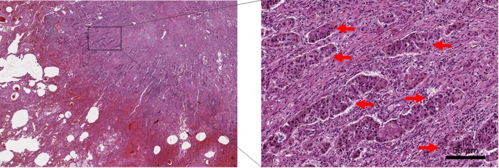
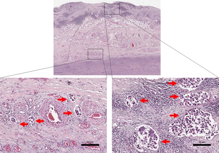
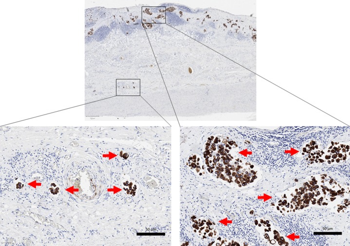
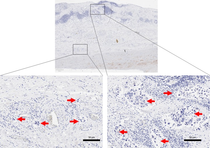
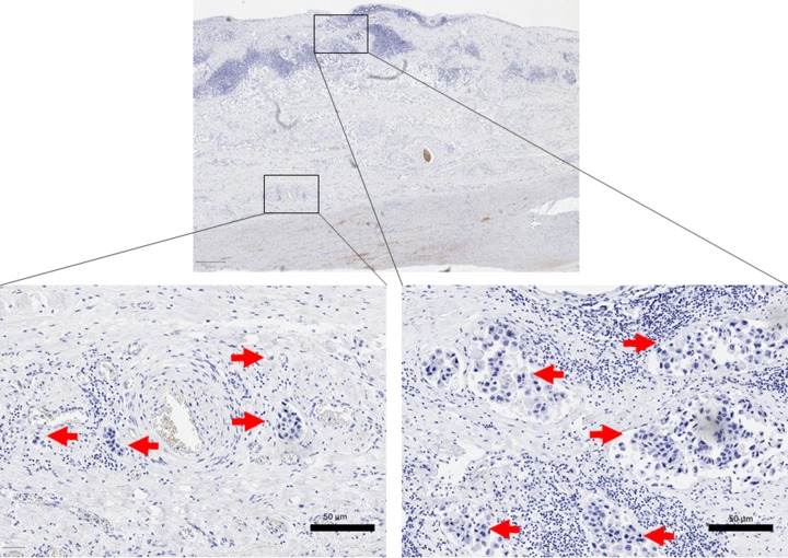
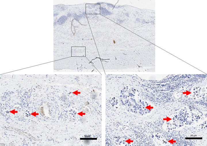
In patients with cardiovascular events, such as myocardial infarction or aortic dissection without known risk factors for cardiovascular disease, neoplastic disease should be considered as a differential diagnosis. In this report, we present a case of a 51-year old man with previously undiagnosed non-small lung cancer leading to fatal cardiovascular complications due to hemovascular spread, diagnosed post-mortem. This case illustrates the value of autopsy in unexpected deaths.
Keywords: Autopsy; NSCLC; cardiovascular complications; cardiovascular disease; hemangiosis carcinomatosa.; non-small cell lung cancer.
Copyright: © 2020, Diaconu et al. and Applied Systems.
Conflict of interest statement
Conflict of interests: The authors declare that there is no conflict of interest.
Figures
References
-
- Aortic Tumors. Restrepo Carlos S., Betancourt Sonia L., Martinez-Jimenez Santiago, Gutierrez Fernando R. Seminars in Ultrasound, CT and MRI. 2012;33(3):265-272. - PubMed
-
- Dissection of the ascending aorta due to metastatic carcinoma. Ugurlu B S, Hazan E, Badak O, Yörükoğlu K, Oto O. The Annals of thoracic surgery. 2001;72(2):614–5. - PubMed
-
- Aortic intramural hematoma associated with metastatic carcinoma. Tsuchida Rikuhei, Kasahara Naoto, Inobe Megumi, Terado Yuichi, Horita Ayako, Yokoyama Kenichi, Sakamoto Atsushiko, Fujioka Yasunori, Kurata Atsushi. Pathology, research and practice. 2010;206(12):839–45. - PubMed
LinkOut - more resources
Full Text Sources