Review
. 2020 Mar;6(1):106-123.
doi: 10.21037/jss.2020.01.10.

A review of cervical spine alignment in the normal and degenerative spine

Affiliations
Review

A review of cervical spine alignment in the normal and degenerative spine

Parthik D Patel et al. J Spine Surg. 2020 Mar.

Abstract

With recent advancements in surgical spine technology and techniques, the importance of regional and global spine alignment has become an important factor in surgical planning. Our review aims to consolidate the current literature on cervical and global alignment parameters and its relationship to cervical symptomatology, quality of life (QOL), requirements for surgery, potential surgical complications and health-related quality of life (HRQOL) outcomes.

Keywords: Quality of life (QOL); cervical; complications; global alignment; spine alignment.


Conflict of interest statement

Conflicts of Interest: The series “Advanced Techniques in Complex Cervical Spine Surgery” was commissioned by the editorial office without any funding or sponsorship. The authors have no conflicts of interest to declare.

Figures

Figure 1
Figure 1
Three different methods to determine CL. (A) Left: a, CA—measured as the angle subtended by the line parallel to the inferior endplate of C2 and line parallel to the inferior endplate of C7; (B) middle: b, JPA—measured as the angle between the line parallel to the posterior surface of C2 and C7; (C) right: HPA—measured as the sum of each cervical segmental angle by drawing lines parallel to the posterior surface of C2 to C7. CL, cervical lordosis; CA, Cobb angle; JPA, Jackson physiologic stress; HPA, Harrison posterior angle.
Figure 2
Figure 2
Regional cervical alignment parameters. (A) Left: a, CBVA; b, CCA; c, C0–C2 angle; d, C2–C7 CA (CL); (B) right: e, T1S; f, C2–C7 SVA (cSVA); g, SLS; h, McGS. CBVA, chin-brow vertical angle; CCA, craniocervical angle; CA, Cobb angle; T1S, T1 slope; cSVA, cervical sagittal vertical axis; SLS, slope of line of sight; McGS, McGregor line of slope.
Figure 3
Figure 3
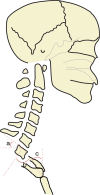
Cervical malalignment with respect to the thoracic inlet. a, T1S; b, TIA; c, NT. T1S, T1 slope; TIA, thoracic inlet angle; NT, neck tilt.
Figure 4
Figure 4
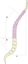
Global spine alignment parameters. a, TK; b, LL; c, SVA. TK, thoracic kyphosis; LL, lumbar lordosis; SVA, sagittal vertical axis.
Figure 5
Figure 5
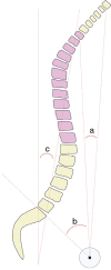
Sacropelvic alignment parameters. a, SS; b, PT; c, PI. SS, sacral slope; PT, pelvic tilt; PI, pelvic incidence.
Figure 6
Figure 6
Lower extremity alignment parameters in the setting of spinal malalignment. a, KFA; b, PS. KFA, knee flexion angle; PS, pelvic shift.
Figure 7
Figure 7
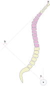
Global spine alignment parameters in the setting of adult spinal deformity. a, CTPA; b, TPA; c, T1-L5 SSI. CTPA, cervico-thoracic pelvic angle; TPA, T1-pelvic angle; SSI, sagittal spinal inclination.
Figure 8
Figure 8
Cervical and global spine alignment parameters. a, C2T; b, C2PT; c, PT. C2T, C2 tilt; C2PT, C2-pelvic tilt; PT, pelvic tilt.

References

    1. Dubousset J. Three-dimensional analysis of the scoliotic deformity. Pediatr Spine Princ Pract 1994.
    1. Ganau M, Zewude R, Fehlings MG. Functional anatomy of the spinal cord. In: Kaiser MG, Haid RW, Shaffrey CI, et al. editors. Degenerative cervical myelopathy and radiculopathy. Cham: Springer International Publishing, 2019:3-12.
    1. Jun BY. Anatomic study for ideal and safe posterior C1-C2 transarticular screw fixation. Spine (Phila Pa 1976) 1998;23:1703-7. 10.1097/00007632-199808010-00018 - DOI - PubMed
    1. Bell GH, Dunbar O, Beck JS, et al. Variations in strength of vertebrae with age and their relation to osteoporosis. Calcif Tissue Res 1967;1:75-86. 10.1007/BF02008077 - DOI - PubMed
    1. Macintosh JE, Bogduk N. The morphology of the lumbar erector spinae. Spine (Phila Pa 1976) 1987;12:658-68. 10.1097/00007632-198709000-00004 - DOI - PubMed