Review
. 2020 Mar;6(1):145-155.
doi: 10.21037/jss.2019.12.01.

Occipital fixation techniques and complications

Affiliations
Review

Occipital fixation techniques and complications

Mohamed Macki et al. J Spine Surg. 2020 Mar.

Abstract

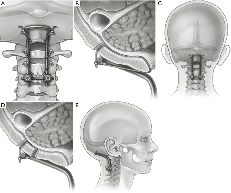
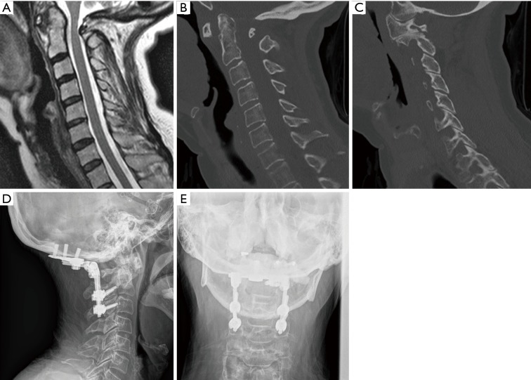
Occipitocervical fusions in the adult population are most commonly indicated for neoplastic tumors invading the craniocervical junction (CCJ), rheumatological deformities compromising the foramen magnum, and traumatic dislocations resulting in occiput-C1 instability. Appropriate preoperative imaging will not only assist in identifying the pathology but also determine a treatment regimen for the diseased junction. A treatment algorithm for craniocervical disease is proposed. Lesions must first be identified as irreducible versus reducible: restore extension and/or distraction of the craniovertebral junction without injuring the neural elements. Irreducible lesions require decompression only, while reducible lesions require an added fusion. Techniques in fusion are broadly divided into external immobilization versus internal fixation. The former entails halo rings and tongs for a prolonged duration. Fixation surgeries vary from wiring to screw fixation of the occiput-C1 segment. Details of the operation as well as potential complications are discussed.

Keywords: Cervical; craniocervical; craniovertebral; occipital; occipitocervical.


Conflict of interest statement

Conflicts of Interest: The series “Advanced Techniques in Complex Cervical Spine Surgery” was commissioned by the editorial office without any funding or sponsorship. V Chang receives research funding from Medtronic, who was not involved in this project, specifically. He is also a consultant for Globus Medical, K2M, and SpineGuard. The other authors have no conflicts of interest to declare.

Figures

Figure 1
Figure 1
The Hartshill-Ransford contoured loop is secured to the suboccipital region in 1 of 2 ways: the wires are threaded one burr hole rostrally and the foramen magnum caudally (A,B), or the wires are threaded through burr holes on each side of the rod (C,D,E). (Figure published with permission from the Journal of Neurosurgery: Pediatrics. Occipitocervical fusion using a contoured rod and wire construct in children: a reappraisal of a vintage technique. Klimo P Jr, Astur N, Gabrick K, et al. J Neurosurg Pediatr 2013;11:160-9. February).
Figure 2
Figure 2
Case presentation: a patient with a history of rheumatoid arthritis presented with progressive neck pain with bilateral upper extremity pain and paresthesias, decline in fine motor skills, arm weakness, bladder incontinence, and gait instability. The patient was positive for Lhermitte’s sign and 3+ brisk deep tendon reflexes. (A) preoperative MRI shows cord compression; (B) preoperative CT shows C1-2 instability and subluxation; (C) preoperative CT shows unfavorable C1 lateral mass anatomy, thus C1-2 fusion is not feasible; (D) anteroposterior and (E) lateral X-ray demonstrating occipitocervical fusion—elective C1 laminectomy was performed.
Figure 3
Figure 3
In the technique described by Faure et al., two occipital hooks are placed back-to-back in the occipitocervical fusion construct. (Figure published with permission from Journal of Neurosurgery. Inverted-hook occipital clamp system in occipitocervical fixation. Faure A, Monteiro R, Hamel O, et al. J Neurosurg 2002;97:135-41. July).

References

    1. Panjabi M, Dvorak J, Crisco J, 3rd, et al. Flexion, extension, and lateral bending of the upper cervical spine in response to alar ligament transections. J Spinal Disord 1991;4:157-67. 10.1097/00002517-199106000-00005 - DOI - PubMed
    1. Dvorak J, Schneider E, Saldinger P, et al. Biomechanics of the craniocervical region: the alar and transverse ligaments. J Orthop Res 1988;6:452-61. 10.1002/jor.1100060317 - DOI - PubMed
    1. Simmons ED, Zheng Y. Vertebral tumors: surgical versus nonsurgical treatment. Clin Orthop Relat Res 2006;443:233-47. 10.1097/01.blo.0000198723.77762.0c - DOI - PubMed
    1. Boriani S, Bandiera S, Biagini R, et al. Chordoma of the mobile spine: fifty years of experience. Spine (Phila Pa 1976) 2006;31:493-503. 10.1097/01.brs.0000200038.30869.27 - DOI - PubMed
    1. Schellinger KA, Propp JM, Villano JL, et al. Descriptive epidemiology of primary spinal cord tumors. J Neurooncol 2008;87:173-9. 10.1007/s11060-007-9507-z - DOI - PubMed