Review
. 2020 Mar;6(1):164-180.
doi: 10.21037/jss.2020.03.05.

Posterior atlantoaxial fusion: a comprehensive review of surgical techniques and relevant vascular anomalies

Affiliations
Review

Posterior atlantoaxial fusion: a comprehensive review of surgical techniques and relevant vascular anomalies

Qi Chen et al. J Spine Surg. 2020 Mar.

Abstract

Posterior atlantoaxial fusion is an important surgical technique frequently used to treat various pathologies involving the cervical 1-2 joint. Since the beginning of the 20th century, various fusion techniques have been developed with improved safety profile, higher fusion rates, and superior clinical outcome. Despite the advancement of technology and surgical techniques, posterior C1-2 fusion is still a technically challenging procedure given the complex bony and neurovascular anatomy in the craniovertebral junction (CVJ). In addition, vascular anomalies in this region are not uncommon and can lead to devastating neurovascular complications if unrecognized. Thus, it is important for spine surgeons to be familiar with various posterior atlantoaxial fusion techniques along with a thorough knowledge of various vascular anomalies in the CVJ. Intimate knowledge of the various surgical techniques in combination with an appreciation for anatomical variances, allows the surgeon develop a customized surgical plan tailored to each patient's particular pathology and individual anatomy. In this article, we aim to provide a comprehensive review of existing posterior C1-2 fusion techniques along with a review of common vascular anomalies in the CVJ.

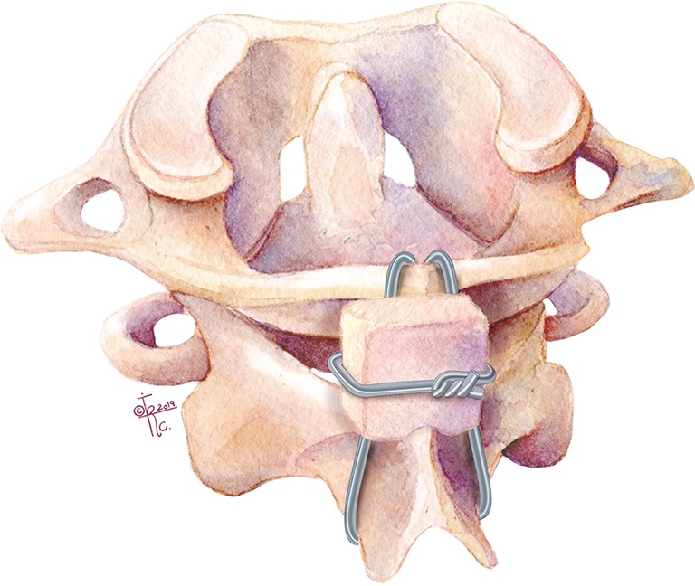
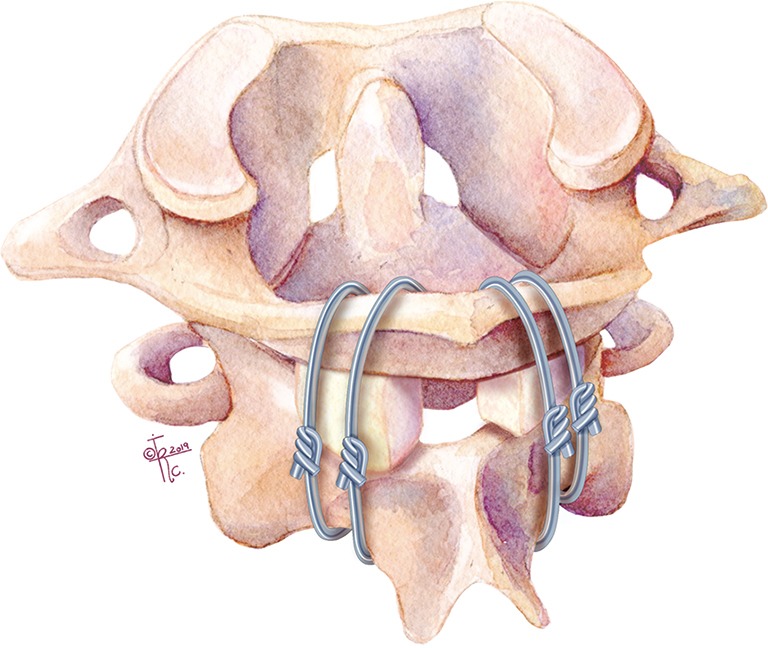
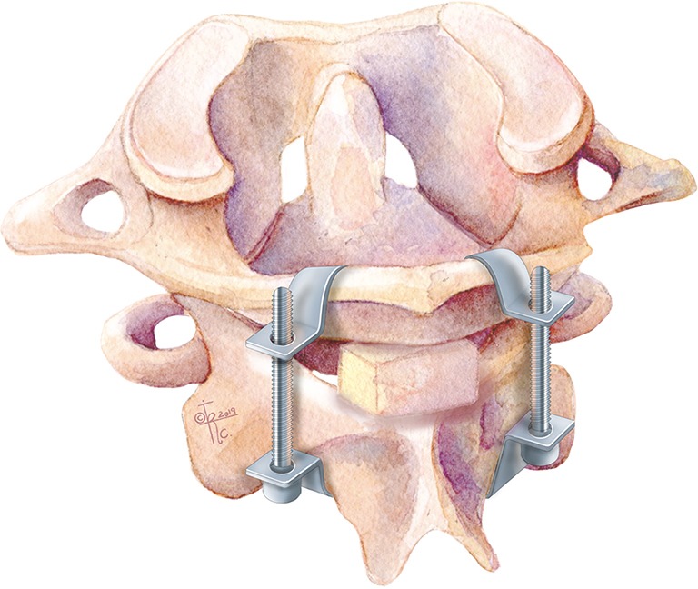
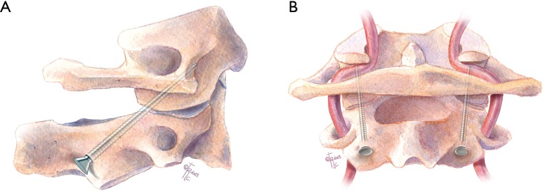
Keywords: Atlantoaxial fusion; Gallie technique; Goel technique; Harm’s technique; Sonntag and Dickman technique; brooks technique; clamp; lateral mass screw; pedicle screw; posterior cervical fusion; screw fixation; transarticular; translaminar; wires.


Conflict of interest statement

Conflicts of Interest: The series “Advanced Techniques in Complex Cervical Spine Surgery” was commissioned by the editorial office without any funding or sponsorship. LAT serves as the unpaid editorial board member of Journal of Spine Surgery from Jan. 2019 to Jan. 2021 and served as the unpaid Guest Editor of the series. The other authors have no conflicts of interest to declare.

Figures

Figure 1
Figure 1
Illustration of the Gallie sublaminar technique. Wire placed under C1 posterior arch, looping around the C2 spinous process, and then performing “on-lay” fusion by placing iliac crest bone with a midline notch to dock onto the C2 lamina and spinous process.
Figure 2
Figure 2
Illustration of the Brooks and Jenkins sublaminar technique. Modification of the Gallie technique by placing two separate pieces of iliac crest bone graft between C1 posterior arch and C2 lamina. In addition, there is a separate sublaminar wire under both C1 and C2 laminae on each side.
Figure 3
Figure 3
Illustration of Halifax technique (interlaminar clamps).
Figure 4
Figure 4
Illustration of transarticular screw (TAS). Threaded screws inserted into C2 pars ending in the C1 lateral masses. (A) Sagittal and (B) coronal planes.
Figure 5
Figure 5
Illustration of Goel and Laheri technique. Posterior C1 lateral mass-C2 pars screws connected by posterior cervical plates.
Figure 6
Figure 6
Illustration of the Harms technique. Modification of the Goel technique in which C1 lateral mass screws are connected to C2 pedicle screws by rods. (A) Sagittal and (B) coronal planes.
Figure 7
Figure 7
Illustration of “pedicle” screw fixation. Insertion of C1 lateral mass screw via the posterior arch. (A) Axial and (B) sagittal planes.
Figure 8
Figure 8
Illustration of C1 posterior arch crossing screw technique. Insertion of crossing screw into the C1 posterior arch.
Figure 9
Figure 9
Illustration of C2 intralaminar screw fixation. Insertion of translaminar screws as fixation points at C2 to connect with C1 lateral mass screws.
Figure 10
Figure 10
Illustration of C1 hook combined with C2 pedicle screw.
Figure 11
Figure 11
Example of case in which C1 lateral mass and C2 pedicle screw fixation with sublaminar wiring were used. (A) Sagittal CT of pre-reduction os odontoideum; (B) parasagittal CT of C1–C2 fixation using lateral mass and pars screws; (C) sagittal CT of post-reduction os odontoideum; (D) axial CT demonstrating pars screw placement in C2.

References

    1. Nakajima K, Onomura T, Tanida Y, et al. Factors related to the severity of myelopathy in atlantoaxial instability. Spine (Phila Pa 1976) 1996;21:1440-5. 10.1097/00007632-199606150-00008 - DOI - PubMed
    1. Ringel F, Reinke A, Stuer C, et al. Posterior C1-2 fusion with C1 lateral mass and C2 isthmic screws: accuracy of screw position, alignment and patient outcome. Acta Neurochir (Wien) 2012;154:305-12. 10.1007/s00701-011-1224-x - DOI - PubMed
    1. Brooks AL, Jenkins EB. Atlanto-axial arthrodesis by the wedge compression method. J Bone Joint Surg Am 1978;60:279-84. 10.2106/00004623-197860030-00001 - DOI - PubMed
    1. Gallie WE. Skeletal traction in the treatment of fractures and dislocations of the cervical spine. Ann Surg 1937;106:770-6. 10.1097/00000658-193710000-00026 - DOI - PMC - PubMed
    1. Goel A. Atlantoaxial joint jamming as a treatment for atlantoaxial dislocation: a preliminary report. Technical note. J Neurosurg Spine 2007;7:90-4. 10.3171/SPI-07/07/090 - DOI - PubMed