

Anatomic techniques for cervical pedicle screw placement
- PMID: 32309664
- PMCID: PMC7154374
- DOI: 10.21037/jss.2020.03.07
Anatomic techniques for cervical pedicle screw placement
Abstract
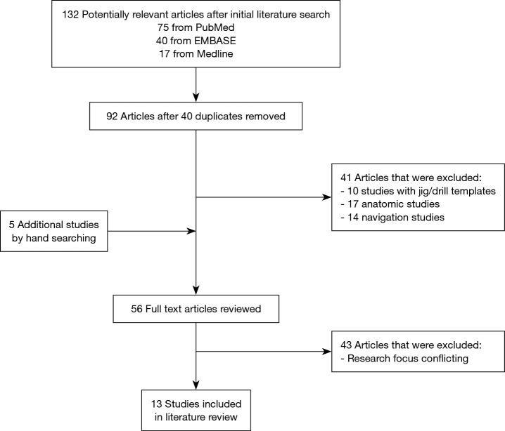
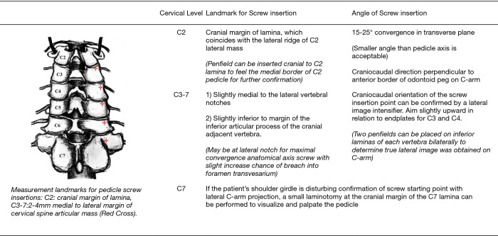
Instrumentation of the cervical spine with cervical pedicle screws (CPS) is beneficial in patients with various types of spinal pathology. Despite posing greater technical challenges, CPS instrumentation confers better fixation outcomes when compared to lateral mass screws. While developments in technology have augmented the accuracy of CPS insertion, mastery in freehand CPS insertion allows the aforementioned technologies to reach their full potential in improving patient outcomes. The aim of this article is to discuss freehand CPS insertion techniques as established in the current literature while sharing our experience in this context. A comprehensive literature search was performed using the following electronic databases: PubMed, Medline, and EMBASE. Full-text articles focusing on clinical studies with description of freehand techniques were included. Articles which were on cadaveric studies, drill jig, navigation or robotic technology were excluded. Thirteen primary references comprising 1,480 patients were included in this review. Majority of studies reported utilizing the cranial margin of lamina for C2 level as a landmark for entry point, as well as lateral to centre of the articular mass, and just medial to the lateral border of the superior articular process for C3-7 levels. Method of tracking and facilitation of trajectory was reported in multiple studies, with use of instruments ranging from curved pedicle probes to high-speed burrs. Limited studies reported specific trajectories of CPS insertion. Most studies noted testing pedicle wall integrity at various checkpoints, with pedicle screw repositioning or conversion to lateral screw mass following detection of perforation or screw malpositioning. Success in CPS insertion rests on meticulous preoperative planning to identify the ideal screw entry point and trajectory. Patient-specific drill jigs, navigation and robotic technologies, while beneficial to progress in the field of cervical spine surgery and patient outcomes, should serve primarily to augment good expertise in freehand CPS insertion technique.
Keywords: Cervical spine; insertion; pedicle screw; technique.
2020 Journal of Spine Surgery. All rights reserved.
Conflict of interest statement
Conflicts of Interest: The series “Advanced Techniques in Complex Cervical Spine Surgery” was commissioned by the editorial office without any funding or sponsorship. The authors have no conflicts of interest to declare.
Figures
References
-
- Hojo Y, Ito M, Suda K, et al. A multicenter study on accuracy and complications of freehand placement of cervical pedicle screws under lateral fluoroscopy in different pathological conditions: CT-based evaluation of more than 1,000 screws. Eur Spine J 2014;23:2166-74. 10.1007/s00586-014-3470-0 - DOI - PubMed
Publication types
LinkOut - more resources
Full Text Sources
Miscellaneous