Review
. 2020 Mar;6(1):290-301.
doi: 10.21037/jss.2020.01.05.

Cervical laminoplasty: indication, technique, complications

Affiliations
Review

Cervical laminoplasty: indication, technique, complications

Douglas S Weinberg et al. J Spine Surg. 2020 Mar.

Abstract

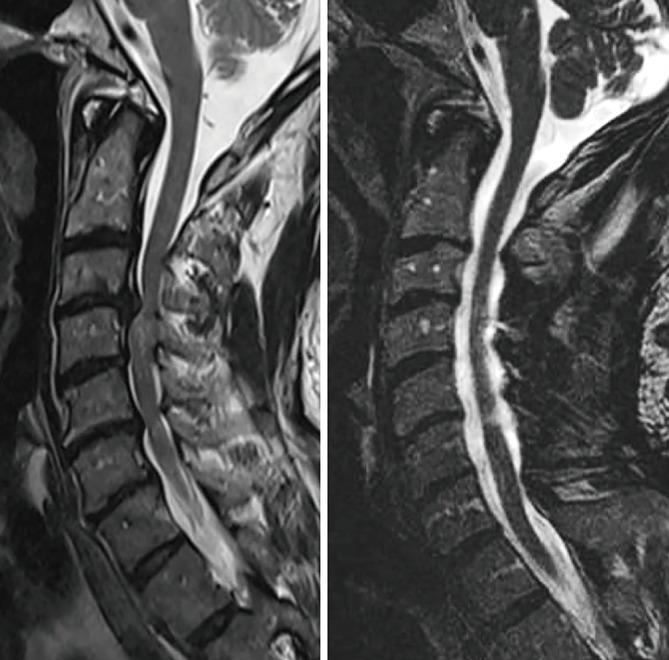
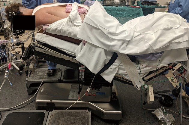
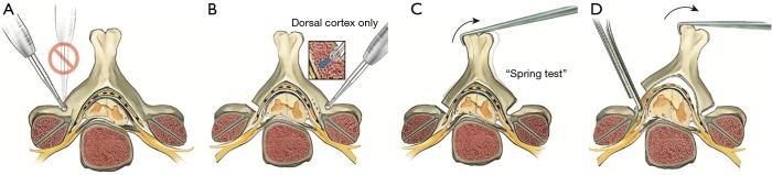
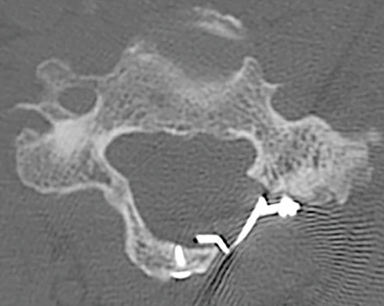
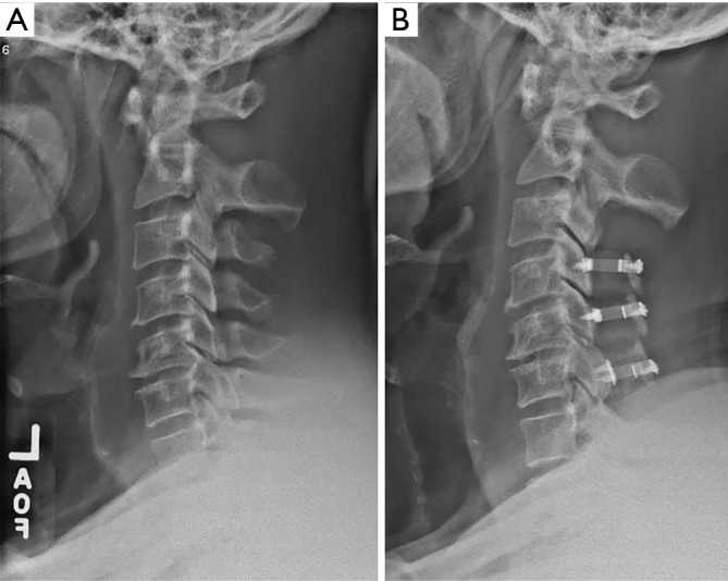
Cervical laminoplasty is a non-fusion, decompression procedure for cervical spondylotic myelopathy (CSM). It is most commonly indicated for patients with multilevel stenosis who have preserved sagittal alignment and minimal to no axial neck pain related to spondylosis. Expansion of the laminar arch can allow for direct and indirect decompression of the spinal canal. Relative contraindications include those patients with significant preoperative neck pain, kyphotic alignment and substantial instability. Potential advantages over laminectomy and fusion include avoiding fusion-related complications, and the preservation of motion. Important technical considerations include meticulous extensor muscle management, with special attention being given to preserving the soft tissue attachments to C2. In the properly selected patient, outcomes are comparable, and in some studies superior, to other operations for CSM.

Keywords: Sagittal balance; cervical myelopathy; cervical spine surgery; cervical stenosis; laminoplasty.


Conflict of interest statement

Conflicts of Interest: The series “Advanced Techniques in Complex Cervical Spine Surgery” was commissioned by the editorial office without any funding or sponsorship. JM Rhee: royalties from Biomet and Stryker; is a member of a speakers’ bureau or has made paid presentations on behalf of Biomet/Zimmer, Medtronic, and Depuy; serves as a paid consultant to Biomet, Synthes; has received research or institutional support from Depuy, Johnson & Johnson Company, Kineflex, and Medtronic; and serves as a board member of the Cervical Spine Research Society. Receives royalties from Wolters-Kluwer. Royalties for a laminoplasty plate. DS Weinberg has no conflicts of interest to declare.

Figures

Figure 1
Figure 1
Preoperative (A) and postoperative (B) T2 sagittal images of a patient with cervical myelopathy treated with laminoplasty. Note the drift-back of the spinal cord away from the anterior impinging structures postoperatively, leading to an indirect decompression of the anterior cord. There has also been a direct decompressive effect from the posterior compressive elements on the dorsal aspect of the cord, and the diameter of the spinal canal has been substantially enlarged.
Figure 2
Figure 2
A properly positioned patient for laminoplasty. The Mayfield head holder is used to position the neck in a neutral or slightly flexed alignment. Care is taken to pad the knees, legs, and abdomen. Note that the patient is placed in reverse Trendelenburg position such that the orientation of the cervical spine is roughly parallel to the floor, with flexion of the knees to ensure the patient does not migrate caudally. The popliteal fossa should be examined to ensure the calf muscles are not excessively tense. The shoulders are taped and the arms are tucked to facilitate soft tissue tension and X-ray acquisition.
Figure 3
Figure 3
Laminoplasty technique. (A) The opening is created at the lateral mass-laminar junction by angling the burr perpendicular to the lamina—towards the spinal canal—rather than vertically into the facet joint; (B) the trough is completed on the opposite side, leaving the ventral cortex intact; (C) greenstick fractures are created by placing dorso-lateral tension on the spinous process or cut edge of lamina; (D) the ligamentum flavum is put under tension and cut with a Kerrison rongeur. Printed with permission from: Saadat et al., Cervical laminoplasty. In: Rhee JM. editor. Emory’s Illustrated Tips and Tricks in Spine Surgery. 1st edition. Wolters Kluwer Health, 2019.
Figure 4
Figure 4
Axial CT scan demonstrating recreation of an expanded laminar arch, as well as bony union on the hinge side.
Figure 5
Figure 5
Preoperative (A) and postoperative (B) lateral X-rays of a patient who underwent C3 laminectomy with C4–6 laminoplasty. Note preservation of preoperative alignment with no substantial change in lordosis or cervical sagittal balance.

References

    1. Fehlings MG, Tetreault LA, Riew KD, et al. A Clinical Practice Guideline for the Management of Patients With Degenerative Cervical Myelopathy: Recommendations for Patients With Mild, Moderate, and Severe Disease and Nonmyelopathic Patients With Evidence of Cord Compression. Global Spine J 2017;7:70S-83S. 10.1177/2192568217701914 - DOI - PMC - PubMed
    1. Veidlinger OF, Colwill JC, Smyth HS, et al. Cervical myelopathy and its relationship to cervical stenosis. Spine (Phila Pa 1976) 1981;6:550-2. 10.1097/00007632-198111000-00004 - DOI - PubMed
    1. Pavlov H, Torg JS, Robie B, et al. Cervical spinal stenosis: determination with vertebral body ratio method. Radiology 1987;164:771-5. 10.1148/radiology.164.3.3615879 - DOI - PubMed
    1. Nagata K, Yoshimura N, Hashizume H, et al. Physical performance decreases in the early stage of cervical myelopathy before the myelopathic signs appear: the Wakayama Spine Study. Eur Spine J 2019;28:1217-24. 10.1007/s00586-019-05907-4 - DOI - PubMed
    1. Tetreault LA, Karadimas S, Wilson JR, et al. The natural history of degenerative cervical myelopathy and the rate of hospitalization following spinal cord injury: an updated systematic review. Global Spine J 2017;7:28S-34S. 10.1177/2192568217700396 - DOI - PMC - PubMed