Review
. 2020 Aug 1;41(4):bnaa007.
doi: 10.1210/endrev/bnaa007.

GDF15: A Hormone Conveying Somatic Distress to the Brain

Affiliations
Review

GDF15: A Hormone Conveying Somatic Distress to the Brain

Samuel M Lockhart et al. Endocr Rev. .

Abstract

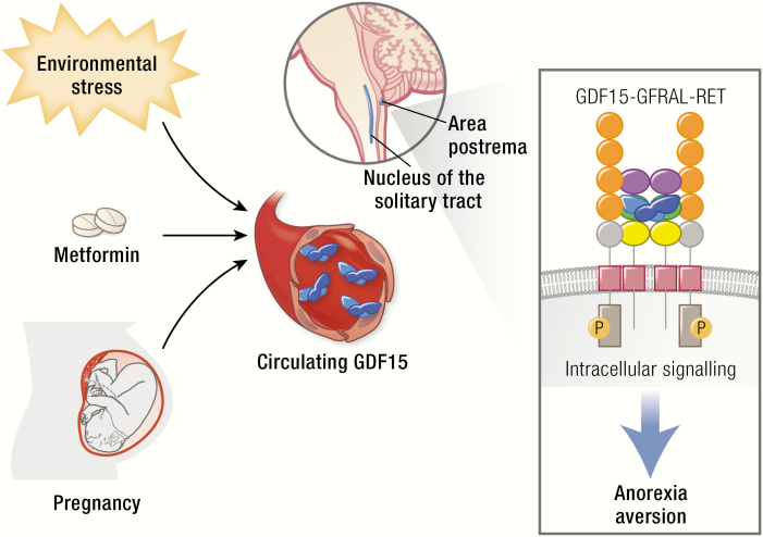
GDF15 has recently gained scientific and translational prominence with the discovery that its receptor is a GFRAL-RET heterodimer of which GFRAL is expressed solely in the hindbrain. Activation of this receptor results in reduced food intake and loss of body weight and is perceived and recalled by animals as aversive. This information encourages a revised interpretation of the large body of previous research on the protein. GDF15 can be secreted by a wide variety of cell types in response to a broad range of stressors. We propose that central sensing of GDF15 via GFRAL-RET activation results in behaviors that facilitate the reduction of exposure to a noxious stimulus. The human trophoblast appears to have hijacked this signal, producing large amounts of GDF15 from early pregnancy. We speculate that this encourages avoidance of potential teratogens in pregnancy. Circulating GDF15 levels are elevated in a range of human disease states, including various forms of cachexia, and GDF15-GFRAL antagonism is emerging as a therapeutic strategy for anorexia/cachexia syndromes. Metformin elevates circulating GDF15 chronically in humans and the weight loss caused by this drug appears to be dependent on the rise in GDF15. This supports the concept that chronic activation of the GDF15-GFRAL axis has efficacy as an antiobesity agent. In this review, we examine the science of GDF15 since its identification in 1997 with our interpretation of this body of work now being assisted by a clear understanding of its highly selective central site of action.

Keywords: GDF15; GFRAL; RET; cachexia; hyperemesis gravidarum; obesity.


Figures

Graphical Abstract
Graphical Abstract
Figure 1.
Figure 1.
GDF15: Gene and protein structure. A: Schematic representation of the human genomic segment containing GDF15 between neighbor genes PGPEP1 and LRCC25. GDF15 in blue (coding exons) and white (UTR); the 3’ and 5’ coding exons of PGPEP1 and LRCC25, respectively, in black; miRNA miR-3189 in yello; and the experimentally established transcription factor binding sites in the promoter for ▲ CHOP, ■ P53, and ● Sp1/Egr1. Sense of the GDF15 transcript is from left to right. The arrows indicate the putative causal SNPs in 5 different haplotypes that might influence GDF15 transcription. The red arrow indicates the rs1054564 variant in the GDF15 3’-UTR that has been experimentally validated to alter GDF15 expression via altered miRNA binding (16). B: Schematic demonstrating the signal peptide (blue), propeptide (orange), and mature peptide of GDF15. Pairing of the 6th cysteine in 2 pro-GDF15 monomers forms a pro-GDF15 dimer, which can be secreted and bound to the extracellular matrix or proteolytic cleavage at an “RXXR” motif and can liberate the mature peptide, which is secreted and circulates as a bioactive homodimer. C: Schema illustrating the cysteine–cysteine pairing within, and between, GDF15 monomers.
Figure 2.
Figure 2.
Evolution of GDF15 sequence and structure compared to TGF-β1. A: Amino acid alignment of GDF15 sequences from vertebrates and human TGF-β1. Selection and alignment are based on all available sequences and a single representative sequence is displayed for placentals, marsupials, monotherms, birds, reptiles, amphibians, coelacanths and fishes. Supplementary Figure 1 available at (31) displays more representative sequences in higher resolution. The bar under the alignment indicates the subdomains identified in the structure of TGF-β1: grey, regions absent in placental GDF15 (ie, the straitjacket helix at the N-terminus and the second part of the arm); blue, the rest of the propeptide; yellow, active ligand TGF-β1; black, conserved, helix-stabilizing motif in the strait jacket. The same color coding of the subdomains is used in the right-hand monomer in Fig. 3B. A large insertion 117–155 in the Gallus gallus sequence is not displayed (blue line). The residues affected by the common coding SNPs are indicated by triangles, H202 in red. B: Structure of human pro-TGFβ-1 (32). The subdomains in the monomer on the right is colored as described in Fig. 2A, the left is in green (propeptide) and magenta (ligand). The broken lines stand for the mobile loops invisible in the crystal structure. Grey color indicates the regions that are missing in the GDF15 of placentalia. The straitjacket is the N-terminal helix (bottom) where the black part indicates the strongly conserved, helix-stabilizing motif aEaaR (notably absent in placentalia—c.f. Fig. 2A) with the side chains of E and R forming a helix-stabilizing ion pair displayed. The position corresponding to the polymorphic H202D in human GDF15 is indicated in red.
Figure 3.
Figure 3.
The GDF15-GFRAL-RET complex. The same color scheme is used to highlight the protein domains in all 3 panels. A: The gene structure of the human GRFAL alongside a schematic domain arrangement of the full-length GFRAL protein and its splice variant isoGFRAL. The coding exons are highlighted in light blue, with the 3’ UTR shown in white. GFRAL domains are labelled signal (signal peptide), D1-3 (GFR-like domain 1-3), TM (transmembrane domain), and cyto (cytoplasmic domain). The splice variant in which exon 6 is skipped as indicated and leading to a premature stop is predicted according to the homologous experimentally established mouse transcript. The resultant isoGFRAL lacks D3 and the downstream transmembrane domain and could function as a soluble GFRAL receptor isoform. B: Schema depicting GDF15, full-length GFRAL, the putative splice variant isoGFRAL, and RET. RET domains are labelled CLD1-4 (Cadherin-Like), CRD (cysteine rich), TM (trans membrane), and KIN (kinase). The activated (phosphorylated) receptor complex GDF15-GFRAL-RET is shown in the middle, while the putative isoGFRAL receptor is depicted extracellularly bound to GDF15. C: CryoEM structure of GDF15-GFRAL-RET ectodomain (PDB access code 6Q2J, surface visualization). The 2 GDF15 molecules in the dimer are distinguished in different shades of blue. Arrows indicate the linkers to the membrane for RET (left) and GFRAL (right) to the membrane (the remaining 2 are at the rear side).

References

    1. Bootcov MR, Bauskin AR, Valenzuela SM, et al. MIC-1, a novel macrophage inhibitory cytokine, is a divergent member of the TGF-beta superfamily. Proc Natl Acad Sci U S A. 1997;94(21):11514–11519. - PMC - PubMed
    1. Lawton LN, Bonaldo MF, Jelenc PC, et al. Identification of a novel member of the TGF-beta superfamily highly expressed in human placenta. Gene. 1997;203(1):17–26. - PubMed
    1. Böttner M, Suter-Crazzolara C, Schober A, Unsicker K. Expression of a novel member of the TGF-beta superfamily, growth/differentiation factor-15/macrophage-inhibiting cytokine-1 (GDF-15/MIC-1) in adult rat tissues. Cell Tissue Res. 1999;297(1):103–110. - PubMed
    1. Paralkar VM, Vail AL, Grasser WA, et al. Cloning and characterization of a novel member of the transforming growth factor-beta/bone morphogenetic protein family. J Biol Chem. 1998;273(22):13760–13767. - PubMed
    1. Hromas R, Hufford M, Sutton J, Xu D, Li Y, Lu L. PLAB, a novel placental bone morphogenetic protein. Biochim Biophys Acta. 1997;1354(1):40–44. - PubMed

Publication types

Substances