. 2020 Aug 19;15(16):1515-1528.
doi: 10.1002/cmdc.202000160. Epub 2020 May 6.

Pentathiepins: A Novel Class of Glutathione Peroxidase 1 Inhibitors that Induce Oxidative Stress, Loss of Mitochondrial Membrane Potential and Apoptosis in Human Cancer Cells

Affiliations

Pentathiepins: A Novel Class of Glutathione Peroxidase 1 Inhibitors that Induce Oxidative Stress, Loss of Mitochondrial Membrane Potential and Apoptosis in Human Cancer Cells

Steven Behnisch-Cornwell et al. ChemMedChem. .

Abstract

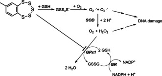
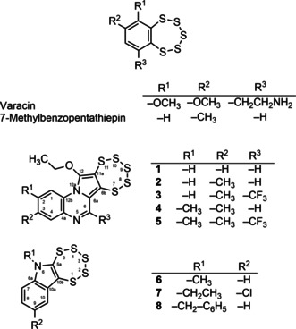
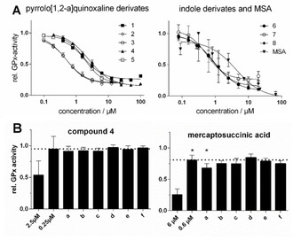
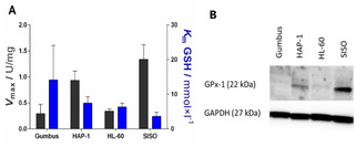
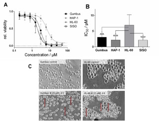
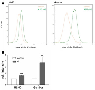
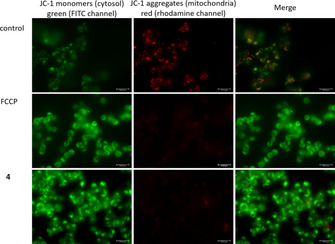
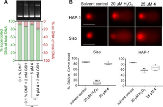
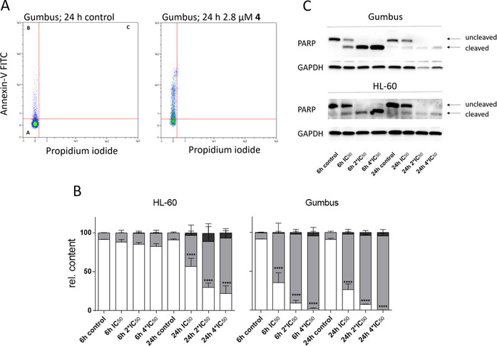
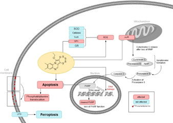
A novel class of glutathione peroxidase 1 (GPx1) inhibitors, namely tri- and tetracyclic pentathiepins, has been identified that is approximately 15 times more potent than the most active known GPx1 inhibitor, mercaptosuccinic acid. Enzyme kinetic studies with bovine erythrocyte GPx1 indicate that pentathiepins reversibly inhibit oxidation of the substrate glutathione (GSH). Moreover, no inhibition of superoxide dismutase, catalase, thioredoxin reductase or glutathione reductase was observed at concentrations that effectively inhibit GPx1. As well as potent enzyme inhibitory activity, the pentathiepins show strong anticancer activity in various human cancer cell lines, with IC50 values in a low-micromolar range. A representative tetracyclic pentathiepin causes the formation of reactive oxygen species in these cells, the fragmentation of nuclear DNA and induces apoptosis via the intrinsic pathway. Moreover, this pentathiepin leads to a rapid and strong loss of mitochondrial membrane potential in treated cancer cells. On the other hand, evidence for the induction of ferroptosis as a form of cell death was negative. These new findings show that pentathiepins possess interesting biological activities beyond those originally ascribed to these compounds.

Keywords: DNA fragmentation; apoptosis; cancer cells; cytotoxicity; glutathione peroxidase; oxidative stress; pentathiepin.


Conflict of interest statement

The authors declare no conflict of interest.

Figures

Figure 1
Figure 1
Postulated mechanism for the oxidative DNA damaging activity of 7‐methylbenzopentathiepin16, 17 and the role that GSH and GPx1 inhibition could play in this activity. GSH: reduced glutathione; GSSG: glutathione disulfide; GSSxS: polysulfide anions; SOD: superoxide dismutase; GR: glutathione reductase.
Figure 2
Figure 2
Structures and systematic numbering of pentathiepins.
Scheme 1
Scheme 1
Synthesis of pentathiepins 15 by the molybdenum mediated diethoxy alkyne to pentathiepin route.
Figure 3
Figure 3
Molecular structures of 5 (left) and 8 (right), as determined by X‐ray structural analysis. Ellipsoids are drawn at the 50 % probability level.
Scheme 2
Scheme 2
Synthesis of pentathiepins 68 by the disulfur dichloride route.
Figure 4
Figure 4
A) Concentration‐activity plots of bovine GPx1, inhibited by various pentathiepins and MSA at 23 °C (mean±SD); B) Residual relative GPx activity determined directly at the noted inhibitor concentrations or (a–f) after a 100‐fold jump‐dilution of incubations at the higher inhibitor concentration (i. e., 2.5 and 6.0 μM), pre‐incubated for 30 min at 23 °C under the following conditions: a) GPx (5 U/L)+inhibitor+250 μM GSH+500 μM t‐BHO; b) GPx (5 U/L)+inhibitor+250 μM GSH; c) GPx (5 U/L)+inhibitor; d) inhibitor+250 μM GSH+500 μM t‐BHO; e) Inhibitor+250 μM GSH; f) inhibitor alone. The dotted line represents the enzyme activity expected after the jump‐dilution if inhibition were to be 100 % reversible. *) p<0.01, two‐sided, paired t‐test, n=5.
Figure 5
Figure 5
A) GPx activity of cancer cell lysates expressed as maximum velocity per mg [U/mg] (black) and corresponding Michaelis‐Menten constant (K m; blue; mean±SD, n>3) at 23 °C. B) Western blot analyses of human GPx1 in various cancer cell lines.
Figure 6
Figure 6
A) Relative cell viability after exposure to various concentrations of 4 for 48 h (mean±SD, n>3); B) IC50 values for the reduction of cell viability of 4 in various cell lines (mean±confidence interval 95 %, n=3; ***p<0.001; ****p<0.0001; C) Morphology of Gumbus and HL‐60 cells after exposure to 25 μM 4 for 6 h (induced membrane blebbing highlighted by arrows).
Figure 7
Figure 7
A) Representative flow cytometric histograms of ROS determination with DCF‐DA‐labeled HL‐60 (left) and Gumbus (right) cells after incubation with 25 μM 4 for 10 min (green) or vehicle control (orange) at λ em/ex=488/530 nm, B) Relative increase of ROS after incubation with 25 μM 4 for 10 min (mean+SD, n>4, ***p<0.001, ****p<0.0001).
Figure 8
Figure 8
Fluorescence microscopy of HAP‐1 cells after incubation with FCCP or 2.2 μM 4 for 1 h followed by staining with JC‐1 dye; Left: JC‐1 monomers in cytosol, emission of green light (FITC channel); middle: JC 1 aggregates in mitochondria, emission of red light (rhodamine channel); right: overlay.
Figure 9
Figure 9
A) Supercoiled plasmid cleavage by compound 4 or the respective solvent DMF in the absence and presence of GSH, displayed as ratios of supercoiled (left y‐axis) and open circular (right y‐axis) plasmid DNA. Data from three independent replicates were compared by two‐tailed unpaired t‐test (n=3; **p<0.01). B) Above: Representative images of comets in HAP‐1 and SISO cells exposed to either 1 % DMF as solvent, H2O2 or 4 for 15 min at 0 °C. Scale bar: 50 μm. Below: Box and‐whiskers plot displaying the percentage of intact genomic DNA in the comet head after various treatments. One‐way ANOVA and Dunnett's multiple comparisons tests were performed relative to the solvent control (n≥3; *p<0.05; **p<0.01; ****p<0.0001).
Figure 10
Figure 10
A) Representative flow cytometric plots of Annexin‐V/PI‐labeled Gumbus cells, with and without exposure to 2.8 μM 4 for 24 h (population in quadrant I: living cells; II: apoptotic cells; III: late apoptotic/necrotic cells), B) Results of cytometric studies with Annexin‐V/PI‐labeled HL‐60 and Gumbus cells treated with 4 at multiple concentrations of the viability IC50 for 6 or 24 h; living cells (white), early apoptotic cells (gray), late apoptotic/necrotic cells (black; mean+SD, n=3, ****p<0.0001); C) Detection of PARP cleavage by western blotting in Gumbus and HL‐60 cells after incubation with various concentrations of 4 for 6 or 24 h.
Figure 11
Figure 11
Summary of the proposed biological effects of pentathiepin 4 in cancer cells. Red:affect, blue: not affected

References

    1. Lubos E., Loscalzo J., Handy D. E., Antioxid. Redox Signaling 2011, 15, 1957–1997. - PMC - PubMed
    1. Rocher C., Lalanne J. L., Chaudiere J., Eur. J. Biochem. 1992, 205, 955–960. - PubMed
    1. Toppo S., Flohe L., Ursini F., Vanin S., Maiorino M., Biochim. Biophys. Acta 2009, 1790, 1486–1500. - PubMed
    1. None
    1. Ho Y. S., Magnenat J. L., Bronson R. T., Cao J., Gargano M., Sugawara M., Funk C. D., J. Biol. Chem. 1997, 272, 16644–16651; - PubMed

MeSH terms