Review
. 2020 Aug;21(8):459-474.
doi: 10.1038/s41580-020-0236-x. Epub 2020 Apr 20.

The regulation and functions of DNA and RNA G-quadruplexes

Affiliations
Review

The regulation and functions of DNA and RNA G-quadruplexes

Dhaval Varshney et al. Nat Rev Mol Cell Biol. 2020 Aug.

Abstract

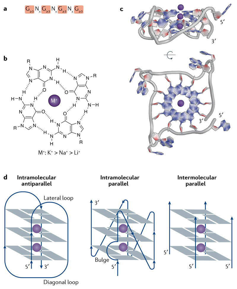
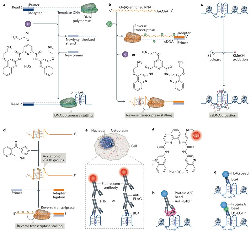
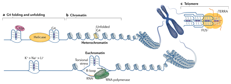
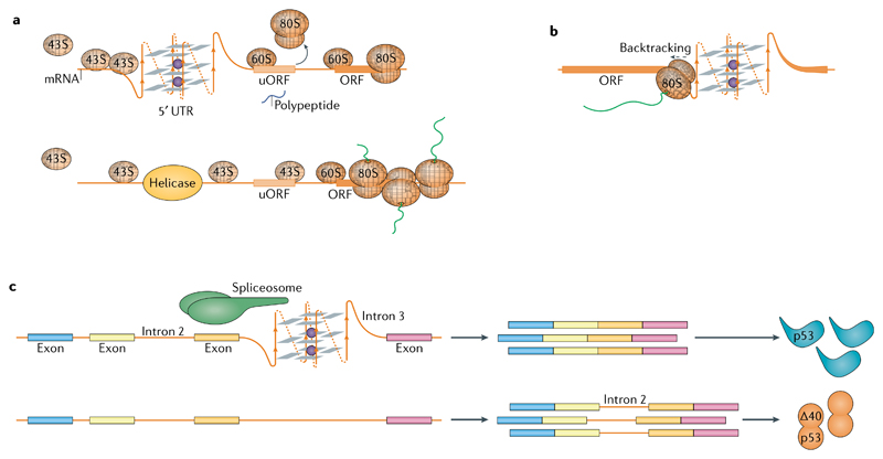
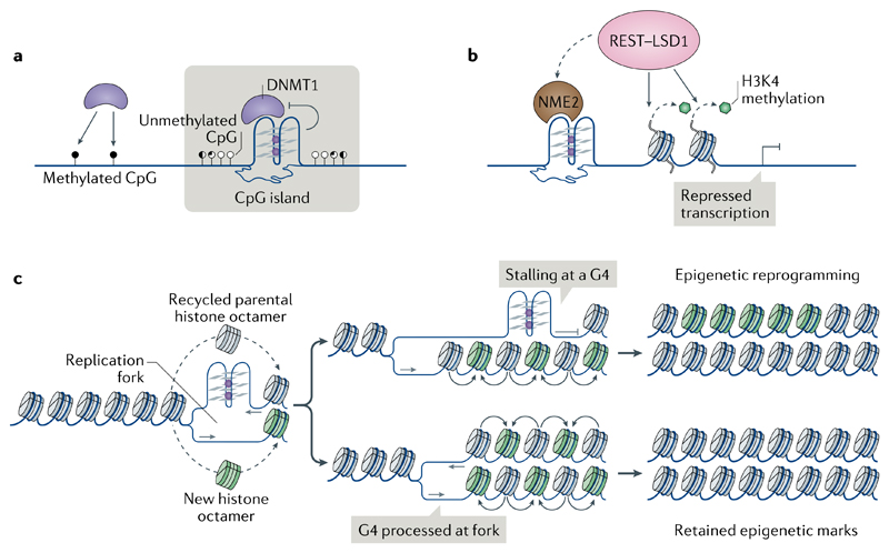
DNA and RNA can adopt various secondary structures. Four-stranded G-quadruplex (G4) structures form through self-recognition of guanines into stacked tetrads, and considerable biophysical and structural evidence exists for G4 formation in vitro. Computational studies and sequencing methods have revealed the prevalence of G4 sequence motifs at gene regulatory regions in various genomes, including in humans. Experiments using chemical, molecular and cell biology methods have demonstrated that G4s exist in chromatin DNA and in RNA, and have linked G4 formation with key biological processes ranging from transcription and translation to genome instability and cancer. In this Review, we first discuss the identification of G4s and evidence for their formation in cells using chemical biology, imaging and genomic technologies. We then discuss possible functions of DNA G4s and their interacting proteins, particularly in transcription, telomere biology and genome instability. Roles of RNA G4s in RNA biology, especially in translation, are also discussed. Furthermore, we consider the emerging relationships of G4s with chromatin and with RNA modifications. Finally, we discuss the connection between G4 formation and synthetic lethality in cancer cells, and recent progress towards considering G4s as therapeutic targets in human diseases.


Conflict of interest statement

Competing interests

S.B. is a founder and shareholder of Cambridge Epigenetix Ltd.

Figures

Fig. 1
Fig. 1. The structure and topologies of G-quadruplexes.
a | The G-quadruplex (G4) consensus sequence. x denotes the number of nucleotides in the loops (see part d). b | A guanine tetrad is stabilized by Hoogsteen base-pairing and by a central cation (M+), with a preference for monovalent cations in the order of potassium (K+) > sodium (Na+) > lithium (Li+). c | X-ray crystal structure of an intramolecular, parallel G4 from a human telomere sequence (PDB: 1KF1). d | Schematic representation of some G4 topologies.
Fig. 2
Fig. 2. Approaches to detect and map DNA and RNA G-quadruplexes.
a,b | Mapping G-quadruplexes (G4s) by chain-extension stalling followed by high-throughput sequencing (G4-seq). G4s formed in the genome are mapped using G4-seq and G4s formed in the transcriptome are mapped using an analogous approach with reverse transcriptase stalling (rG4-seq). a | In G4-seq, a library of fragmented genomic DNA is sequenced twice, first in non-G4-forming conditions (Read 1) to provide a reference and then in G4-stabilizing conditions (for example, in the presence of K+ or the G4-stabilizing ligand pyridostatin (PDS; Read 2)) to determine the positions of G4-dependent DNA polymerase stalling. b | In rG4-seq, poly(A)-enriched RNA is reverse transcribed in the presence of Li+ (a non-G4-forming condition) as a reference and with K+ or PDS to map RNA G4-dependent reverse transcriptase stalling. c,d | Chemical methods for mapping G4s. DNA G4s are mapped by potassium permanganate (KMnO4)–single-strand nuclease (S1 nuclease) footprinting and RNA G4s are mapped by selective 2′-OH acylation analysed by primer extension (SHAPE). c | In KMnO4–S1 nuclease footprinting, KMnO4 selectively oxidizes and traps single-stranded DNA (ssDNA), thereby allowing its digestion by S1 nuclease. Subsequent computational analyses infer the formation of DNA G4s based on the nuclease footprints. d | SHAPE utilizes differences in acylation kinetics of RNA 2′-hydroxyl groups treated with 2-methylnicotinic acid imidazolide (NAI) and the ability of these groups to stall reverse transcription, to determine the formation of RNA G4s. e | Visualization of G4s (red foci) by immunofluorescence in the nucleus and cytoplasm using G4 antibodies (for example, the IgG antibody 1H6 or the single-chain variable fragment antibody (ScFv) BG4) together with fluorescently conjugated secondary or tertiary antibodies. f | PhenDC3 is an example of a fluorescence-labelled G4-targeting ligand. g | Mapping of DNA G4s using chromatin immunoprecipitation and next-generation sequencing (ChIP-seq) with G4-specific ScFv antibodies. BG4 precipitates DNA G4 structures from chromatin isolated using G4 ChIP-seq, whereas D1 is used in a ChIP-seq-like approach by expressing the antibody in cells. h | DNA G4 formation can be inferred indirectly by mapping the location of G4-binding proteins (G4BPs) using ChIP-seq.
Fig. 3
Fig. 3. Regulation of G-quadruplex structure formation.
a | Physical factors, such as the presence of stabilizing cations (bottom), length and sequence composition (not shown) of the loops and flanking sequences, determine the thermodynamic stability of G-quadruplexes (G4s). In cells, specialized proteins that unwind G4s (for example, helicases) or bind and stabilize G4s (for example, some G4-binding proteins (G4BPs)) can shape the G4 landscape. b | The genomic structural context, such as the negative superhelicity behind the RNA polymerase complex, as well as the crosstalk with other structural phenomena, like RNA–DNA hybrids known as R-loops that arise from hybridization of the nascent RNA with the template DNA, can contribute to G4 formation (bottom). Chromatin structure seems to have a strong influence on G4 formation, as the majority of endogenous G4s have been mapped to nucleosome-depleted, open chromatin regions. c | Telomere heterochromatin homeostasis can be influenced by the simultaneous binding of proteins, such as fused in sarcoma (FUS), to G4s formed in telomere DNA and in the long non-coding RNA TERRA (telomeric repeat RNA).
Fig. 4
Fig. 4. Models of G-quadruplex involvement in transcription.
a | DNA G-quadruplexes (G4s) upstream of the transcription start site (TSS) or in the gene body could bind or displace transcription factors, resulting in altered transcription. b | During transcription elongation, the separation of DNA strands in the transcription bubble may result in the formation of G4s in gene bodies. G4 formation on the template strand can block the progression of RNA polymerase II (Pol II). c | Gene-body G4s on the non-template strand may facilitate transcription re-initiation. Conversely, such G4s may favour the stable association of nascent RNA (orange) with the template DNA, resulting in the formation of RNA–DNA hybrids known as R-loops and in Pol II stalling. d | Formation of DNA–RNA hybrid G4s between the non-template DNA and the nascent RNA can lead to premature transcription termination.
Fig. 5
Fig. 5. G-quadruplexes in RNA biology.
a | Formation of RNA G-quadruplexes (G4s) impedes scanning of the 5′ untranslated region (UTR) by 43S ribosomes and leads to translation initiation at an upstream open reading frame (uORF) at the expense of translation of the main ORF (top). Helicases, such as DHX36 or DHX9, resolve the G4s and facilitate translation of the main ORF (bottom). b | The 80S ribosomes engaged in translation elongation stall 6–7 nucleotides prior to a G4 within the ORF. Stalling can cause ribosome backtracking and synthesis of an alternative peptide. c | Recognition of RNA G4s by spliceosome-associated RNA-binding proteins directs splicing of nearby introns, for example the second intron of p53.
Fig. 6
Fig. 6. The involvement of G-quadruplexes in epigenetic control.
a | Binding to G-quadruplexes (G4s) inactivates DNA (cytosine-5)-methyltransferase 1 (DNMT1), thereby contributing to hypomethylation at CpG islands. b | Promoter G4s and their associated proteins, such as non-metastatic 2 (NME2), recruit the RE1-silencing transcription factor (REST)–lysine-specific histone demethylase 1A (LSD1) repressor complex to remove the gene-activating methylation of histone H3 Lys4 (H3K4) and repress gene expression. c | Stalling of DNA replication forks at G4s (for example, owing to impaired activity of helicases or the presence of G4-stabilizing ligands) may impair histone recycling through the formation of a post-replicative gap (top). Parental histones with their established modifications (grey) are lost and replaced with new histones with no or with different modifications (green), resulting in local epigenetic reprogramming.

References

    1. Bang I. Untersuchungen über die Guanylsäure. Biochemische. 1910;26:293–311.
    1. Gellert M, Lipsett MN, Davies DR. Helix formation by guanylic acid. Proc Natl Acad Sci USA. 1962;48:2013–2018. - PMC - PubMed
    1. Sen D, Gilbert W. Formation of parallel four-stranded complexes by guanine-rich motifs in DNA and its implications for meiosis. Nature. 1988;334:364–366. [ This paper is an early demonstration of a G4 comprising stacked tetrads with interconnecting loop sequences performed using chemical mapping and providing biological insight. ] - PubMed
    1. Burge S, Parkinson GN, Hazel P, Todd AK, Neidle S. Quadruplex DNA: sequence, topology and structure. Nucleic Acids Res. 2006;34:5402–5415. - PMC - PubMed
    1. Kwok CK, Merrick CJ. G-Quadruplexes: prediction, characterization, and biological application. Trends Biotechnol. 2017;35:997–1013. - PubMed

Publication types