

doi: 10.1590/0100-3984.2018.0042.
Hip pain in childhood
Affiliations
- PMID: 32313339
- PMCID: PMC7159046
- DOI: 10.1590/0100-3984.2018.0042
Item in Clipboard
Hip pain in childhood
Radiol Bras.
2020 Jan-Feb.
Abstract
Hip pain in a child can have infectious, inflammatory, traumatic, neoplastic, or developmental causes, which can make the diagnosis challenging. Meticulous history taking and a detailed clinical examination guide the radiological investigation. In this article, we address some of the main causes of hip pain in childhood and their findings on diagnostic imaging.
Keywords: Adolescent; Arthritis, juvenile; Child; Child, preschool; Hip dislocation, congenital; Hip joint; Pain/etiology.
Figures
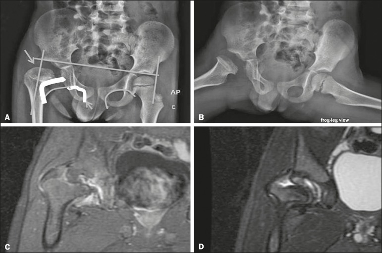
Dysplasia of the right hip in a 4 year-old male. Anteroposterior and frog-leg view X-rays (A and B, respectively), showing asymmetry of the hip joints, with right hip dysplasia. MRI of the right hip. Gadolinium-enhanced coronal, fat-suppressed T1-weighted sequence (C) and STIR sequence (D) showing a loss of sphericity, flattening, and superolateral subluxation of the right femoral head, with enlargement of the femoral neck and acetabular cavity, as well as joint effusion with signs of reactive synovitis.
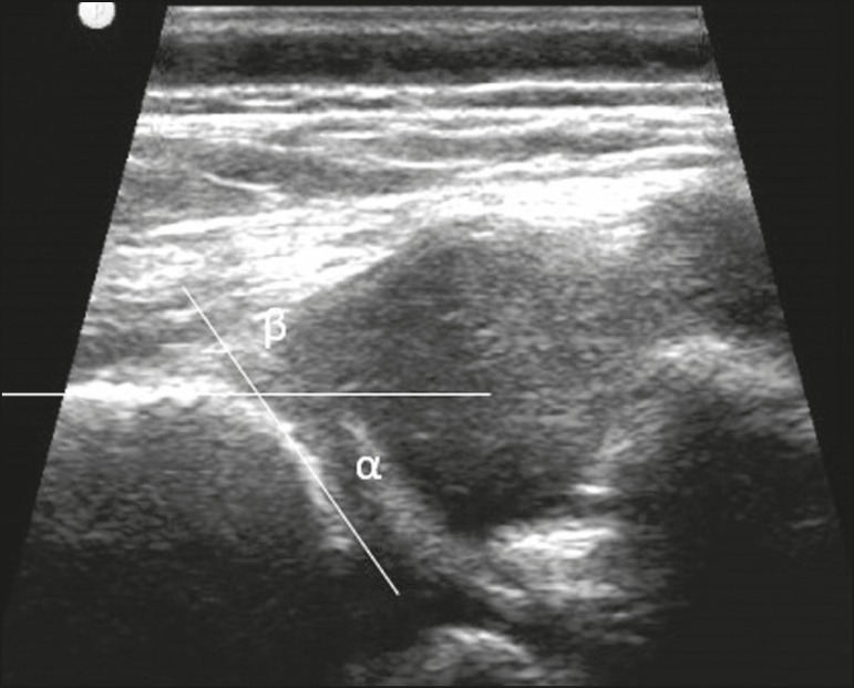
A 40-day-old child presenting left hip dysplasia, characterized by a alpha angle < 60º.
Osteomyelitis in a 9-year-old female. MRI of the right thigh. Coronal fat-suppressed T2-weighted sequence (A) and axial T1-weighted sequence (B) identifying an area in the marrow of the right femoral neck and extending to adjacent muscle planes, with a hypointense signal on the T1-weighted sequence, a hyperintense signal on the T2-weighted sequence, a mild periosteal reaction, and no erosion of cortical bone.
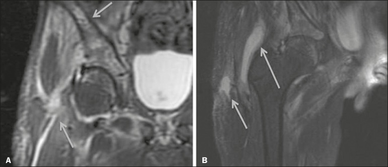
Pyomyositis in a 12-year-old male patient. Coronal fat-saturated T2-weighted MRI sequence showing joint effusion, osteitis, and edema involving the muscle-adipose planes of the gluteal region and root of the right thigh (A), featuring a voluminous, heterogeneous liquid collection, containing small foci of low signal intensity, likely of an infectious/inflammatory nature, located between the ventral portion of the gluteus medius muscle and that of the gluteus maximus muscle (B).
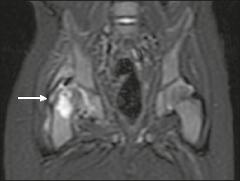
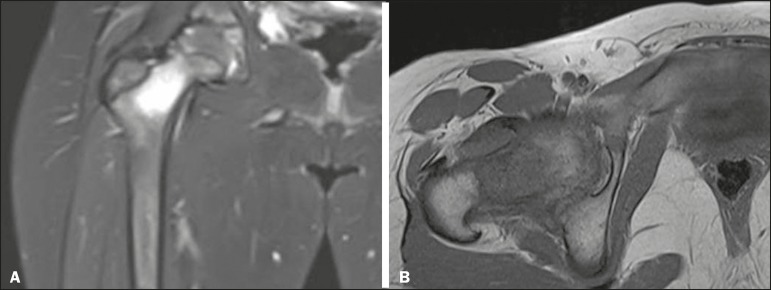
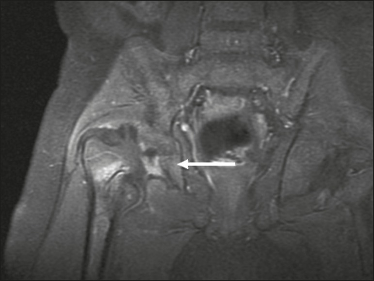
MRI of the hip. STIR coronal sequence showing mild effusion in the right hip joint, with synovial thickening and hyperintense signal/edema in the adjacent planes of the capsule of the muscle. Note the irregular lesion, with a hyperintense signal in a T2-weighted sequence, in the right femoral head and neck, which is suggestive of a focus of osteomyelitis/intraosseous abscess.
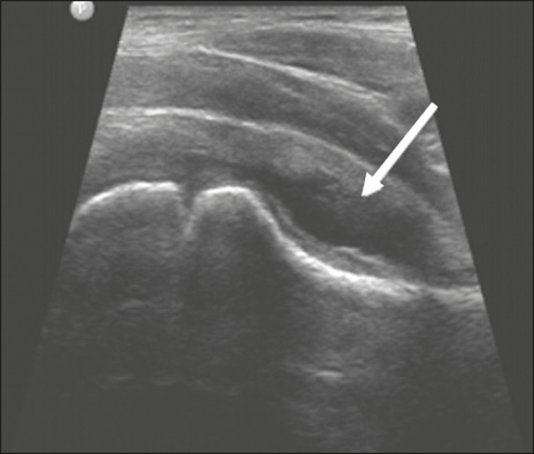
Ultrasound of the hip identifying joint effusion.
A 12-year-old male patient. Sagittal fat-saturated T2-weighted MRI sequence showing moderate intra-articular effusion in the right hip joint.
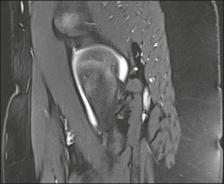
MRI of the hip. STIR coronal sequence showing signs of pronounced epiphysiolysis. Note the moderate joint effusion.
MRI of the hip. Contrast-enhanced, coronal, fat-saturated T1-weighted sequence showing complete destruction/resorption of the femoral head, accompanied by severe deformity of the femoral neck and acetabulum.
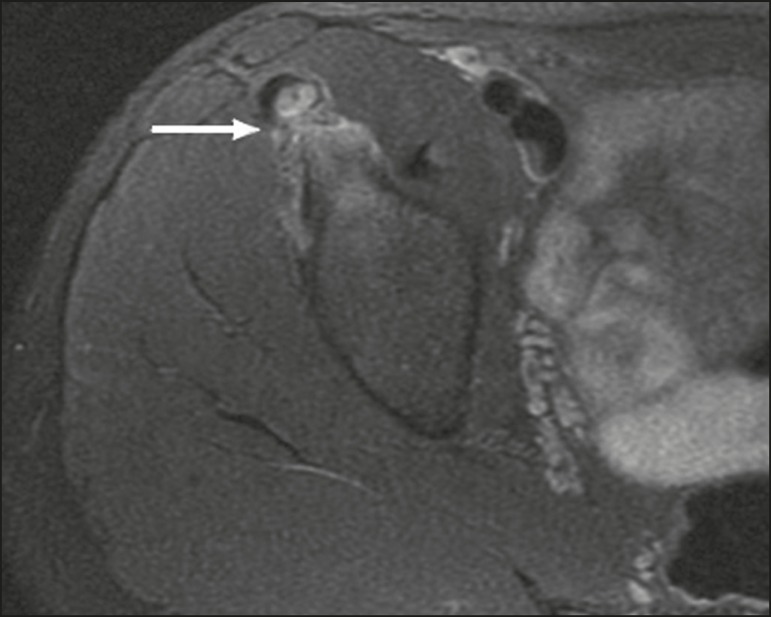
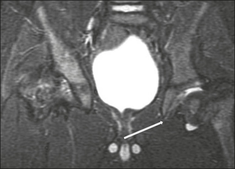
A 14-year-old male. Axial proton density-weighted fat-saturated MRI showing bone edema in the right anteroinferior iliac spine.
References
-
- Zucker EJ, Lee EY, Restrepo R, et al. Hip disorders in children. AJR Am J Roentgenol. 2013;201:W776–W796. - PubMed
LinkOut - more resources
Full Text Sources