Review
. 2020 Jul;7(4):042805.
doi: 10.1117/1.JMI.7.4.042805. Epub 2020 Apr 11.

Virtual clinical trials in medical imaging: a review

Affiliations
Review

Virtual clinical trials in medical imaging: a review

Ehsan Abadi et al. J Med Imaging (Bellingham). 2020 Jul.

Abstract

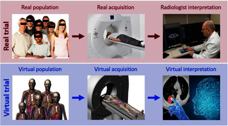
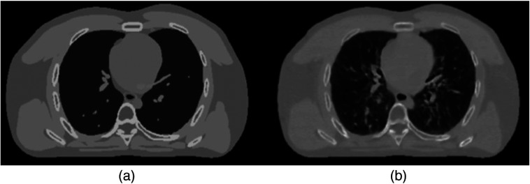
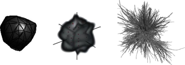
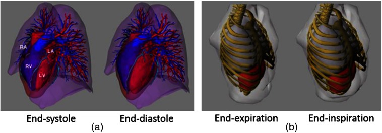
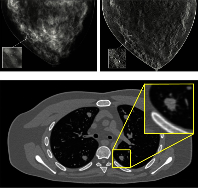
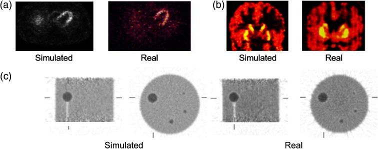
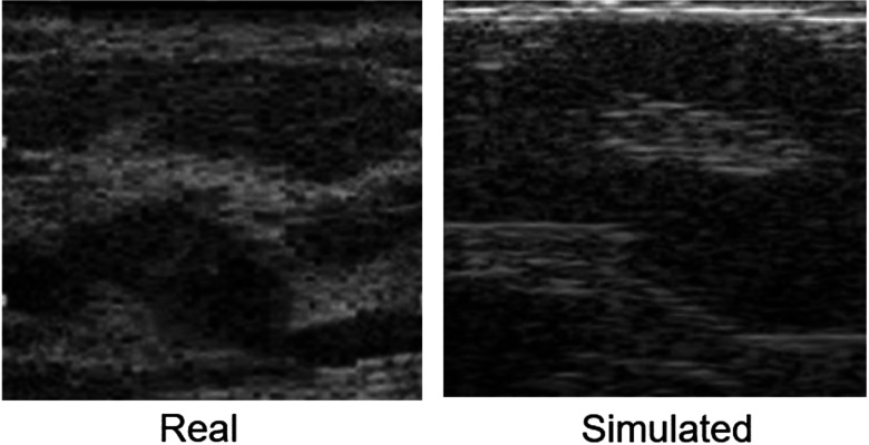
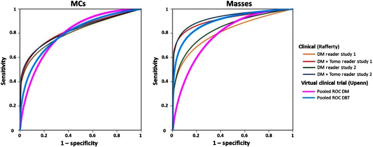
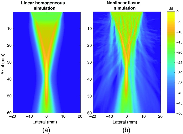
The accelerating complexity and variety of medical imaging devices and methods have outpaced the ability to evaluate and optimize their design and clinical use. This is a significant and increasing challenge for both scientific investigations and clinical applications. Evaluations would ideally be done using clinical imaging trials. These experiments, however, are often not practical due to ethical limitations, expense, time requirements, or lack of ground truth. Virtual clinical trials (VCTs) (also known as in silico imaging trials or virtual imaging trials) offer an alternative means to efficiently evaluate medical imaging technologies virtually. They do so by simulating the patients, imaging systems, and interpreters. The field of VCTs has been constantly advanced over the past decades in multiple areas. We summarize the major developments and current status of the field of VCTs in medical imaging. We review the core components of a VCT: computational phantoms, simulators of different imaging modalities, and interpretation models. We also highlight some of the applications of VCTs across various imaging modalities.

Keywords: computational phantoms; in silico imaging; medical imaging simulation; simulations; virtual clinical trials; virtual imaging trials.


Figures

Fig. 1
Fig. 1
Conducting a clinical imaging trial virtually. Imaging process (top) emulated by virtual imaging trial (bottom).
Fig. 2
Fig. 2
Three main categories of computational phantoms: (a) mathematical based on equations or geometric primitives, (b) voxelized based on segmented imaging data, and (c) BREP based on segmented data but fitting high-level surfaces to the structures. The MIRD, VIP-Man, and XCAT phantoms are shown as examples.
Fig. 3
Fig. 3
Example whole-body phantoms developed by (a) RPI, (b) UF/NCI, (c) IT’IS, and (d) the XCAT series developed by Duke and JHU.
Fig. 4
Fig. 4
Examples of procedurally generated phantoms from UPenn (top) and FDA (bottom).
Fig. 5
Fig. 5
Principal components analysis used for statistically generated phantoms. Top of each breast is simulated mammography projection through all slices. Bottom is a single slice from each phantom.
Fig. 6
Fig. 6
(a) An XCAT phantom lacking intraorgan structures. (b) Same phantom with inserted synthetic textures created by deep learning algorithms.
Fig. 7
Fig. 7
Some examples of simulated lesions using different methodologies. Images adapted from Ref. –.
Fig. 8
Fig. 8
(a) Cardiac motion modeled in the XCAT phantom based on tagged MRI data. (b) RM modeled in the VIP-man based on 4D CT data.
Fig. 9
Fig. 9
Simulated full-field digital mammography (top left), digital breast tomosynthesis (top right), and CT images all with embedded abnormalities (microcalcification cluster on the top and spiculated lesion on the bottom).
Fig. 10
Fig. 10
Comparison of simulated SPECT and PET images with real clinical images. (a) Comparison of transaxial myocardial perfusion SPECT images. (b) Comparable coronal C11-raclopride PET images of the brain. (c) Transverse orthogonal sections through a 20-cm diameter cylindrical phantom with hot spheres supported by plastic rods. Measured data (left) are from a Siemens/CTI ECAT HR+ scanner and simulated data are the same acquisition generated using the ASIM.
Fig. 11
Fig. 11
Real and simulated ultrasound images of breast. The simulations were done by solving a second-order linear wave equation.
Fig. 12
Fig. 12
ROC curves for the detection of microcalcifications and masses in DM and DBT, comparing clinical trial and VCT results.
Fig. 13
Fig. 13
(a) Reference short-axis image of a cardiac phantom compared with (b) k-t PCA and (c) k-t SPARSE reconstruction results. In both reconstructions, the acquired imaging data were undersampled by eightfold to see the effects on reconstruction. The error distribution in k-t PCA can be seen to be more homogeneous.
Fig. 14
Fig. 14
Ultrasound simulations of tissue using (a) linear and (b) nonlinear techniques showing that nonlinear techniques are valuable for understanding the distribution of acoustic energy in the body.

References

    1. Chase J. G., et al. , “Next-generation, personalised, model-based critical care medicine: a state-of-the art review of in silico virtual patient models, methods, and cohorts, and how to validation them,” Biomed. Eng. Online 17(1), 24 (2018). 10.1186/s12938-018-0455-y - DOI - PMC - PubMed
    1. Kainz W., et al. , “Advances in computational human phantoms and their applications in biomedical engineering—a topical review,” IEEE Trans. Radiat. Plasma Med. Sci. 3(1), 1–23 (2019). 10.1109/TRPMS.2018.2883437 - DOI - PMC - PubMed
    1. Xu X. G., “An exponential growth of computational phantom research in radiation protection, imaging, and radiotherapy: a review of the fifty-year history,” Phys. Med. Biol. 59(18), R233–R302 (2014). 10.1088/0031-9155/59/18/R233 - DOI - PMC - PubMed
    1. Hoogendoorn C., et al. , “A high-resolution atlas and statistical model of the human heart from multislice CT,” IEEE Trans. Med. Imaging 32(1), 28–44 (2012). 10.1109/TMI.2012.2230015 - DOI - PubMed
    1. Snyder W. S., et al. , “Estimates of absorbed dose fractions for monoenergetic photon sources uniformly distributed in various organs of a heterogeneous phantom,” J. Nucl. Med. 10(Suppl 3, Pamphlet #5), 7–52 (1969). - PubMed