. 2020 Jun 4;48(10):5749-5765.
doi: 10.1093/nar/gkaa253.

Structural and functional characterization of the severe fever with thrombocytopenia syndrome virus L protein

Affiliations

Structural and functional characterization of the severe fever with thrombocytopenia syndrome virus L protein

Dominik Vogel et al. Nucleic Acids Res. .

Erratum in

Abstract

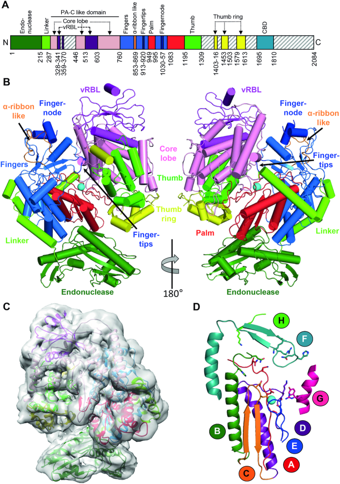
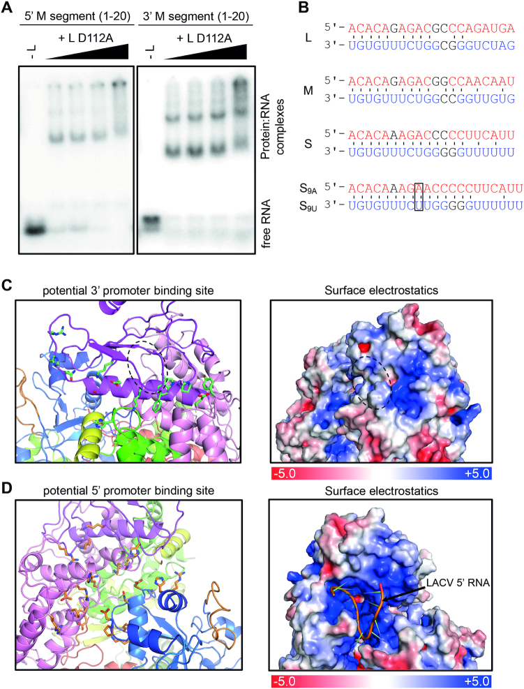
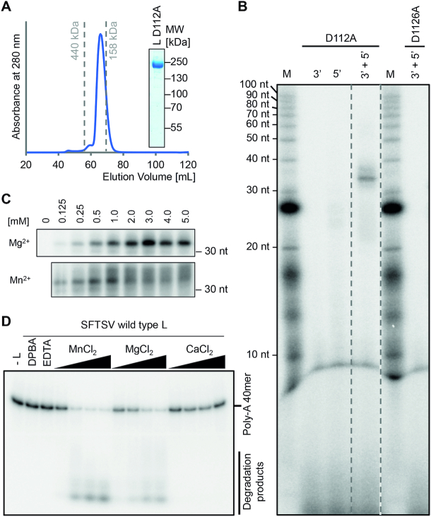
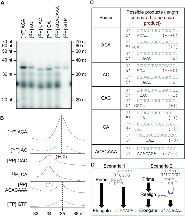
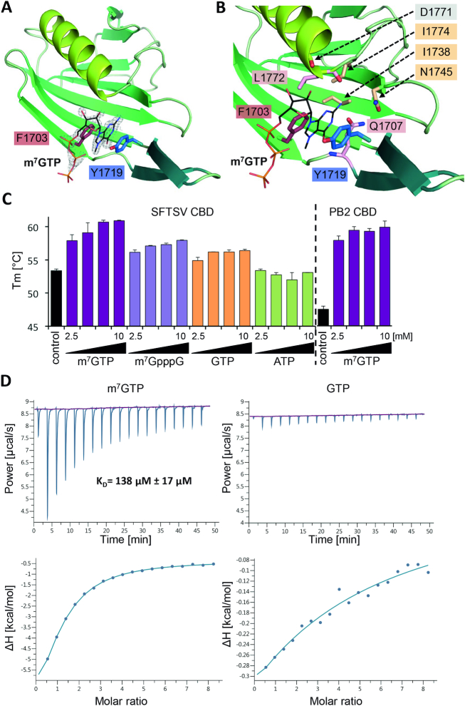
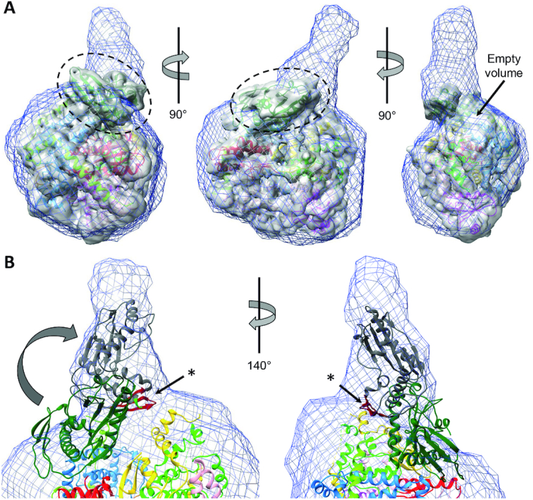
The Bunyavirales order contains several emerging viruses with high epidemic potential, including Severe fever with thrombocytopenia syndrome virus (SFTSV). The lack of medical countermeasures, such as vaccines and antivirals, is a limiting factor for the containment of any virus outbreak. To develop such antivirals a profound understanding of the viral replication process is essential. The L protein of bunyaviruses is a multi-functional and multi-domain protein performing both virus transcription and genome replication and, therefore, is an ideal drug target. We established expression and purification procedures for the full-length L protein of SFTSV. By combining single-particle electron cryo-microscopy and X-ray crystallography, we obtained 3D models covering ∼70% of the SFTSV L protein in the apo-conformation including the polymerase core region, the endonuclease and the cap-binding domain. We compared this first L structure of the Phenuiviridae family to the structures of La Crosse peribunyavirus L protein and influenza orthomyxovirus polymerase. Together with a comprehensive biochemical characterization of the distinct functions of SFTSV L protein, this work provides a solid framework for future structural and functional studies of L protein-RNA interactions and the development of antiviral strategies against this group of emerging human pathogens.


Figures

Figure 1.
Figure 1.
Apo structure of the SFTSV L protein. (A) Schematic linear representation of the domain structure of the SFTSV L protein. (B) Illustrated representation of two views of the apo-L cryo-EM structure as a ribbon diagram (PDB 6Y6K). Domains are colored as in (A). The magnesium ion in the active site is shown as a sphere. A more detailed view and comparison with the LACV L (PDB 5AMQ) and the influenza virus polymerase complex (PDB 6QCV) are shown in Supplementary Figures S2–S4. (C) Superposition of the 5 Å low-pass filtered cryo-EM map with the SFTSV apo-L structure model. (D) Representation of the polymerase core of the SFTSV L with the conserved motifs A–H as a ribbon diagram. The divalent magnesium ion is shown as a sphere.
Figure 2.
Figure 2.
Interaction of the SFTSV L protein with its promoter RNAs. (A) Binding of SFTSV L protein to the 5′ and 3′ promoter ends (20 nt) of the M segment (Supplementary Table S1) was determined by an electrophoretic mobility shift assay. Increasing amounts of L protein (0–1.4 μM) were incubated with 0.2 μM of the indicated RNA (Supplementary Table S1). The protein–RNA complex was separated from the free RNA by native PAGE and visualized by phosphor screen autoradiography using a Typhoon scanner (GE Healthcare). (B) Conserved 3′ (red) and 5′ (blue) terminal sequences of the L, M and S segments. Watson-Crick base pairing is indicated by black lines. Bases which differ between but are conserved within the segments are shown in black. Additional bases at position 9, found in some sequences (5′ S9A, 3′ S9U), are marked with a frame. Potential 3′ (C) and 5′ (D) promoter RNA binding sites are shown as cartoon representation (left). Domains are colored according to Figure 1. Residues potentially involved in protein-RNA interaction are shown as green (C) or orange (D) sticks. See also Supplementary Alignment File. Right panels display surface electrostatics of the potential 3′ and 5′ RNA binding sites generated by APBS within PyMol. LACV 5′ vRNA is shown as a cartoon within the SFTSV potential 5′ vRNA binding site. For comparison, analogous figures for LACV vRNA binding sites are presented in Supplementary Figure S7.
Figure 3.
Figure 3.
In vitro enzymatic activities of the SFTSV L protein. (A) Size exclusion chromatography and Coomassie stained SDS-PAGE analysis of purified SFTSV L (D112A) protein display the high purity and monodispersity of the L protein. Elution volumes of standard proteins (with sizes of 440 and 158 kDa) for column calibration are indicated. (B) The SFTSV L protein mediates RNA synthesis in an in vitro polymerase assay. SFTSV L (D112A) or an RdRp catalytically inactive mutant (D1126A) were incubated with the conserved 5′ or/and 3′ terminal 20 nt of the M segment (5′ M: HO-ACACAGAGACGGCCAACAAU-OH, 3′ M: HO-UGUGUUUCUGGCCGGUUGUG-OH, Supplementary Table S1) in the presence of NTPs supplemented with [α]32P- GTP for 60 min at 30°C. RNA products were separated by denaturing gel electrophoresis and visualized by autoradiography. (C) The RdRp activity of 500 nM SFTSV L (D112A) protein was analyzed in the presence of the indicated concentrations of MgCl2 or MnCl2. (D) 250 nM of wild-type L was incubated with ∼0.3 μM of radioactively labeled PolyA40 RNA substrate (Supplementary Table S1) in the presence of 5, 10, 25 and 50 μM of the indicated Me(II) at 37°C for 30 min. Reactions without L protein, EDTA or the known endonuclease-specific inhibitor DPBA were used as negative controls. Reaction products were separated on a denaturing polyacrylamide gel and visualized by autoradiography.
Figure 4.
Figure 4.
Initiation of replication. (A) RNA products synthesized by the SFTSV L (D112A) protein in the presence of the conserved 5′ and 3′ terminal 20 nt of the M segment (5′ M: HO-ACACAGAGACGGCCAACAAU-OH, 3′ M: HO-UGUGUUUCUGGCCGGUUGUG-OH, Supplementary Table S1) and radioactively labeled primers listed in (C). The experiment was performed as described for the standard polymerase assay (see Materials and Methods). Where indicated, radioactively labeled primers were used instead of [α]32P-GTP. (B) The intensity profiles of the gel lanes from (A) were analyzed using ImageJ software (26) and illustrate the size differences between the product bands shown in (A). Sizes were determined by linear regression using the RNA marker (lower X-axis). (C) The table summarizes the RNA oligonucleotides used as primers in this experiment and the possible products with and without realignment. In brackets the length of the product RNA, relative to the de novo product is given. (D) Schematic representation of the possible priming scenarios. Scenario 1 depicts terminal initiation: ATP primes the reaction by binding to the first nucleotide (position +1) of the template and is further elongated without realignment, resulting in a product with the same length as the de novo reaction product (+/−0). Scenario 2 depicts internal initiation and realignment: the reaction is primed internally by binding of the first ATP to the position +3 and the addition of a C to form a di-nucleotide primer followed by dissociation of the AC dinucleotide and its realignment to position +1 and +2. This realigned AC dinucleotide is then elongated, resulting in the same product as the terminal initiation (+/−0).
Figure 5.
Figure 5.
Structure of SFTSV CBD and m7GTP binding. (A) The figure shows SFTSV CBD crystal structure in complex with an m7GTP. SFTSV CBD is presented as a ribbon diagram with the side chains of the two aromatic residues (F1703, Y1719) involved in stacking interaction with the m7GTP ligand shown as sticks. m7GTP is presented as lines and the surrounding electron density (2|Fo|-|Fc| omit map at 2.0σ) as gray mesh. (B) In a close-up of the m7GTP binding site, the protein is shown as ribbon diagram and side chains involved in m7GTP binding as well as the carbonyl oxygen of residue D1771 are presented as sticks. The m7GTP is shown as lines. The residues I1774, I1738, N1745, are involved in stabilizing the binding site cavity, D1771 (carbonyl oxygen), Q1707 and L1772 directly interact with m7GTP. A detailed list of interactions between the CBD and the m7GTP ligand is given in Supplementary Table S4 and a ligand plot in Supplementary Figure S13A. (C) Thermal stability of SFTSV CBD and influenza A virus PB2 CBD was tested in the presence and absence of different concentrations (2.5, 5.0, 7.5 and 10 mM) of m7GTP, m7GpppG, GTP and ATP. The melting temperatures (Tm) are presented as mean and standard deviations of three independent measurements (n = 3). (D) The affinity of SFTSV CBD for m7GTP and GTP was measured by isothermal titration calorimetry at 25°C. A representative titration curve is shown. Titrations were done three times with 150 μM SFTSV CBD in the cell and 5.0–6.5 mM m7GTP or GTP in the syringe. The upper panel shows the raw data, the lower panel the integrated data fitted to a single-site binding model with the stoichiometry fixed to 1. The dissociation constant KD is given as a mean and standard deviation of three independent measurements for m7GTP.
Figure 6.
Figure 6.
Integrative modelling of SFTSV apo-L protein. (A) A superposition of the SAXS envelope of cluster 1 (blue mesh), a 5Å low-pass filtered cryo-EM mapA (grey surface) and the structure model (ribbon diagram, colored according to Figure 1) is presented in three different orientations. The endonuclease domain, which is sticking out of the SAXS envelope, is marked by a dashed circle. An empty volume is also indicated. (B) Close-up of the endonuclease domain in the superposition of SAXS envelope and structure model. Potential movement of the endonuclease domain is indicated by an arrow and an alternative conformation depicted as grey ribbon diagram. An asterisk indicates the β-sheet (colored in red), for which a role in protein-protein interactions has been proposed.

References

    1. McMullan L.K., Folk S.M., Kelly A.J., MacNeil A., Goldsmith C.S., Metcalfe M.G., Batten B.C., Albarino C.G., Zaki S.R., Rollin P.E. et al. .. A new phlebovirus associated with severe febrile illness in Missouri. N. Engl. J. Med. 2012; 367:834–841. - PubMed
    1. Kohl C., Brinkmann A., Radonic A., Dabrowski P.W., Nitsche A., Muhldorfer K., Wibbelt G., Kurth A.. Zwiesel bat banyangvirus, a potentially zoonotic Huaiyangshan banyangvirus (Formerly known as SFTS)-like banyangvirus in Northern bats from Germany. Sci. Rep. 2020; 10:1370. - PMC - PubMed
    1. Maes P., Alkhovsky S.V., Bao Y., Beer M., Birkhead M., Briese T., Buchmeier M.J., Calisher C.H., Charrel R.N., Choi I.R. et al. .. Taxonomy of the family Arenaviridae and the order Bunyavirales: update 2018. Arch. Virol. 2018; 163:2295–2310. - PubMed
    1. Mehand M.S., Al-Shorbaji F., Millett P., Murgue B.. The WHO R&D Blueprint: 2018 review of emerging infectious diseases requiring urgent research and development efforts. Antiviral Res. 2018; 159:63–67. - PMC - PubMed
    1. Finberg R.W., Lanno R., Anderson D., Fleischhackl R., van Duijnhoven W., Kauffman R.S., Kosoglou T., Vingerhoets J., Leopold L.. Phase 2b study of Pimodivir (JNJ-63623872) as monotherapy or in combination with oseltamivir for treatment of acute uncomplicated seasonal influenza A: TOPAZ trial. J. Infect. Dis. 2019; 219:1026–1034. - PubMed

Publication types