Review
. 2020 Jun;39(2):343-360.
doi: 10.1007/s10555-020-09872-z.

The role of lipid species in membranes and cancer-related changes

Affiliations
Review

The role of lipid species in membranes and cancer-related changes

Tore Skotland et al. Cancer Metastasis Rev. 2020 Jun.

Abstract

Several studies have demonstrated interactions between the two leaflets in membrane bilayers and the importance of specific lipid species for such interaction and membrane function. We here discuss these investigations with a focus on the sphingolipid and cholesterol-rich lipid membrane domains called lipid rafts, including the small flask-shaped invaginations called caveolae, and the importance of such membrane structures in cell biology and cancer. We discuss the possible interactions between the very long-chain sphingolipids in the outer leaflet of the plasma membrane and the phosphatidylserine species PS 18:0/18:1 in the inner leaflet and the importance of cholesterol for such interactions. We challenge the view that lipid rafts contain a large fraction of lipids with two saturated fatty acyl groups and argue that it is important in future studies of membrane models to use asymmetric membrane bilayers with lipid species commonly found in cellular membranes. We also discuss the need for more quantitative lipidomic studies in order to understand membrane function and structure in general, and the importance of lipid rafts in biological systems. Finally, we discuss cancer-related changes in lipid rafts and lipid composition, with a special focus on changes in glycosphingolipids and the possibility of using lipid therapy for cancer treatment.

Keywords: Cancer; Caveolae; Endocytosis; Leaflet interdigitation; Membrane domains; Molecular dynamic simulation.


Figures

Fig. 1
Fig. 1
Illustrations of some lipid structures. Cholesterol is shown on the top followed by PC16:0/16:0, an example of a phospholipid with two saturated fatty acyl chains, which although much used in model membranes is not very common in biological samples. PS 18:0/18:1 is an example of a phospholipid with one saturated and one unsaturated fatty acyl chain, which is a very common combination, and this PS species is the most common PS species in many cells. Note that the unsaturated fatty acyl chains most often are found in the sn-2 position and that all double bonds in phospholipids are in a cis-configuration. PE-P 18:0/20:4 is an example of an ether lipid with an alkenyl chain, i.e., a plasmalogen. The ether lipids often contain polyunsaturated fatty acyl groups in the sn-2 position. Since all double bonds are in the cis-configuration, the polyunsaturated fatty acyl groups will “curl back” towards their head groups and not penetrate into the opposite leaflet even when containing as many carbon atoms as C20:4 (arachidonic acid) or C22:6 (DHA). The sphingolipid SM d18:1/24:1 is shown with the sphingosine part marked in pink. Note that the very long-chain N-amidated fatty acyl chain with 24 carbon atoms is so long that it can reach approximately halfway into the opposite leaflet. Glycosphingolipids contain very little of unsaturated fatty acyl chains, except for the 24:1 that is very common. The structures have been made by using the structure drawing tools available at Lipid Maps (https://lipidmaps.org/)
Fig. 2
Fig. 2
Schematic model of the lipid bilayer of exosomes, which have a lipid composition similar to lipid rafts, e.g., with a high content of SM and CHOL. The number of lipid molecules (excluding CHOL) shown in the outer (29) and inner (21) leaflet is close to the ratio of 1.36 for the outer and inner surface of exosomes with an outer diameter of 70 nm. The lipid composition of the membrane in this simplified illustration is based on the quantitative lipidomic data reported for 22 lipid classes of exosomes excreted from PC-3 cells [20], i.e.,16 SM, 13 PC, 12 PS, 6 PE, 3 PE-O (PE ethers), and 39 molecules of cholesterol (assuming a close to symmetric distribution of cholesterol between the two leaflets). In the right part of the membrane, a possible handshaking between the very-long-chain sphingolipids in the outer leaflet and PS 18:0/18:1 in the inner leaflet in the presence of cholesterol is illustrated. In the rest of the membrane, the lipids are distributed more or less evenly. Nine out of the 16 SM molecules shown contain a very-long-chain N-amidated fatty acyl chain in accordance with the data published. The figure is reproduced from [11]
Fig. 3
Fig. 3
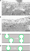
Two EM pictures (a and b) are showing examples of caveolae in cardiac capillary endothelium. These images are reproduced from van Deurs et al. [55] with permission from Elsevier. The scale bar in b is 200 nm. The two drawings at the bottom (c and d) illustrate the possibility for transendothelial transport via caveolae without formation of free intracellular vesicles

References

    1. Devaux PF, Morris R. Transmembrane asymmetry and lateral domains in biological membranes. Traffic. 2004;5(4):241–246. doi: 10.1111/j.1600-0854.2004.0170.x. - DOI - PubMed
    1. van Meer G, Voelker DR, Feigenson GW. Membrane lipids: Where they are and how they behave. Nature Reviews. Molecular Cell Biology. 2008;9(2):112–124. doi: 10.1038/nrm2330. - DOI - PMC - PubMed
    1. Simons K, Toomre D. Lipid rafts and signal transduction. Nature Reviews. Molecular Cell Biology. 2000;1(1):31–39. doi: 10.1038/35036052. - DOI - PubMed
    1. Lingwood D, Simons K. Lipid rafts as a membrane-organizing principle. Science. 2010;327(5961):46–50. doi: 10.1126/science.1174621. - DOI - PubMed
    1. Levental I, Veatch S. The continuing mystery of lipid rafts. Journal of Molecular Biology. 2016;428(24 Pt A):4749–4764. doi: 10.1016/j.jmb.2016.08.022. - DOI - PMC - PubMed

Publication types