Case Reports
. 2020 Jul;92(7):699-702.
doi: 10.1002/jmv.25915.

Central nervous system involvement by severe acute respiratory syndrome coronavirus-2 (SARS-CoV-2)

Affiliations
Case Reports

Central nervous system involvement by severe acute respiratory syndrome coronavirus-2 (SARS-CoV-2)

Alberto Paniz-Mondolfi et al. J Med Virol. 2020 Jul.

Abstract

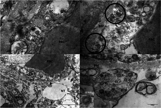
Neurologic sequelae can be devastating complications of respiratory viral infections. We report the presence of virus in neural and capillary endothelial cells in frontal lobe tissue obtained at postmortem examination from a patient infected with severe acute respiratory syndrome coronavirus-2 (SARS-CoV-2). Our observations of virus in neural tissue, in conjunction with clinical correlates of worsening neurologic symptoms, pave the way to a closer understanding of the pathogenic mechanisms underlying central nervous system involvement by SARS-CoV-2.

Keywords: CNS infection; SARS-CoV-2; endothelium; neuroinvasion; neurotropism.


Conflict of interest statement

All the authors declare that there are no conflict of interests.

Figures

Figure 1
Figure 1
A, Brain capillary endothelial cells showing virus particles within cytoplasmic vacuoles (← arrow) B, Blebbing of viral particles coming in/out of the endothelial cell wall (circles) The relationship of virus particles (arrows←) to the endothelial cells (virus ingress/egress) is depicted. Note the dense inner core and densely stained periphery of viral particles. C, Endothelial neural cell interface showing a cytoplasmic vacuole filled with viral particles in various stages of bud formation (arrow←) adjacent to the basement membrane within the neural cell (frontal lobe). D, Neural intracytoplasmic vesicle showing viral‐like particles. Insert: Detail on viral particle exhibiting electron dense centers with distinct stalk‐like peplomeric projections. Scale bars are shown at the bottom left/right of each figure. BM, basement membrane; EC, endothelial cells; IV, intracytoplasmic vesicles; NT, neural tissue; RBC, red blood cell

Comment in

References

    1. Gorbalenya AE, Baker SC, Baric RS, et al. The species severe acute respiratory syndrome‐related coronavirus: classifying 2019‐nCoV and naming it SARS‐CoV‐2. Nat Microbiol. 2020;5:536‐544. 10.1038/s41564-020-0695-z - DOI - PMC - PubMed
    1. Huang C, Wang Y, Li X, et al. Clinical features of patients infected with 2019 novel coronavirus in Wuhan, China. Lancet. 2020;395:497‐506. - PMC - PubMed
    1. Baig AM. Neurological manifestations in COVID‐19 caused by SARS‐CoV‐2. CNS Neurosci Ther. 2020;00:1‐3. - PMC - PubMed
    1. Desforges M, Le Coupanec A, Dubeau P, et al. Human coronaviruses and other respiratory viruses: underestimated opportunistic pathogens of the central nervous system? Viruses. 2019;12:14. - PMC - PubMed
    1. Bohmwald K, Gálvez N, Ríos M, Kalergis AM. Neurologic alterations due to respiratory virus infections. Front Cell Neurosci. 2018;12:386. 10.3389/fncel.2018.00386 - DOI - PMC - PubMed

Publication types

MeSH terms