

[Effects of farnesyltransferase silencing on the migration and invasion of tongue squamous cell carcinoma]
- PMID: 32314892
- PMCID: PMC7184273
- DOI: 10.7518/hxkq.2020.02.012
[Effects of farnesyltransferase silencing on the migration and invasion of tongue squamous cell carcinoma]
Abstract
Objective: This study aimed to explore the effects of silencing farnesyltransferase (FTase) on the migration and invasion of tongue squamous cell carcinoma (TSCC) through RNA interference.
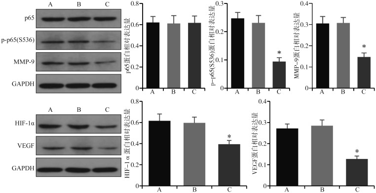
Methods: TSCC cells (CAL27 and SCC-4) were cultured in vitro and then transfected with siRNA to silence FTase expression. The tested cells were categorized as follows: experimental group (three RNA interference groups), negative control group, and blank control group. mRNA expression of FTase and HRAS in each group was analyzed by quantitative real-time polymerase chain reaction. On the basis of FTase mRNA expression, the optimum interference group (highest silencing efficiency) was selected as the experimental group for further study. The protein expression of FTase, HRAS, p65, p-p65(S536), matrix metalloprotein-9 (MMP-9), hypoxia inducible factor-1α (HIF-1α), and vascular endothelial growth factor (VEGF) was analyzed by Western blot. The invasion and migration abilities of TSCC cells were determined by Transwell invasion assay and cell wound healing assay.
Results: The mRNA and protein expression of FTase in the experimental group decreased compared with that in the negative control and blank control groups (P<0.05). The mRNA and protein expression of HRAS was not significantly different among the groups (P>0.05). In the experimental group, the protein expression of p-p65(S536), MMP-9, HIF-1α, and VEGF decreased (P<0.05), whereas that of p65 had no significant change (P>0.05). The migration and invasion abilities of the experimental group were inhibited significantly (P<0.05).
Conclusions: Silencing FTase in vitro could effectively downregulate its expression in TSCC cell lines and reduce the migration and invasion abilities to a certain extent. FTase could be a new gene therapy target of TSCC, and this research provided a new idea for the clinical treatment of TSCC.
目的 通过RNA干扰沉默法尼基转移酶(FTase),探讨沉默FTase对舌鳞状细胞癌迁移和侵袭的影响及其相关机制。方法 针对FTase设计并构建3条小干扰RNA(siRNA),转染人舌鳞状细胞癌CAL27和SCC-4细胞(实验组),同时设置阴性对照组(转染NC-siRNA)和空白对照组(不转染siRNA)。应用实时荧光定量PCR检测各组细胞FTase、HRAS的mRNA表达,根据FTase mRNA的表达量,选取沉默效率最高的FTase-siRNA转染组作为进一步研究的实验组。蛋白质免疫印迹法检测各组细胞FTase、HRAS、p65、p-p65(S536)、基质金属蛋白酶9(MMP-9)、低氧诱导因子1α(HIF-1α)、血管内皮生长因子(VEGF)的蛋白表达,Transwell侵袭实验和细胞划痕实验检测各组细胞的侵袭和迁移能力。结果 与阴性对照组和空白对照组相比,实验组FTase的mRNA和蛋白表达下降(P< 0.05),HRAS的mRNA和蛋白表达无明显变化(P>0.05),p-p65(S536)、MMP-9、HIF-1α、VEGF蛋白表达下降(P<0.05),p65蛋白表达无明显变化(P>0.05);实验组的细胞侵袭和迁移能力降低(P<0.05)。结论 体外沉默舌鳞状细胞癌细胞FTase,可以抑制细胞体外迁移和侵袭能力,为FTase可能作为舌癌治疗的分子靶点提供了一定的理论和实验依据.
Keywords: farnesyltransferase; invasion; migration; tongue squamous cell carcinoma.
Conflict of interest statement
利益冲突声明:作者声明本文无利益冲突。
Figures


References
-
- Jing Y, Jin Y, Wang YJ, et al. SPARC promotes the proliferation and metastasis of oral squamous cell carcinoma by PI3K/AKT/PDGFB/PDGFRβ axis[J] J Cell Physiol. 2019;234(9):15581–15593. - PubMed
MeSH terms
Substances
LinkOut - more resources
Full Text Sources
Research Materials
Miscellaneous