. 2020 Jun 22;60(6):3277-3286.
doi: 10.1021/acs.jcim.0c00179. Epub 2020 May 4.

Fast Identification of Possible Drug Treatment of Coronavirus Disease-19 (COVID-19) through Computational Drug Repurposing Study

Affiliations

Fast Identification of Possible Drug Treatment of Coronavirus Disease-19 (COVID-19) through Computational Drug Repurposing Study

Junmei Wang. J Chem Inf Model. .

Abstract

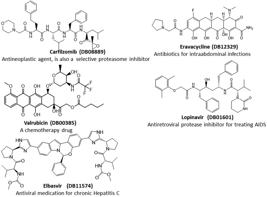
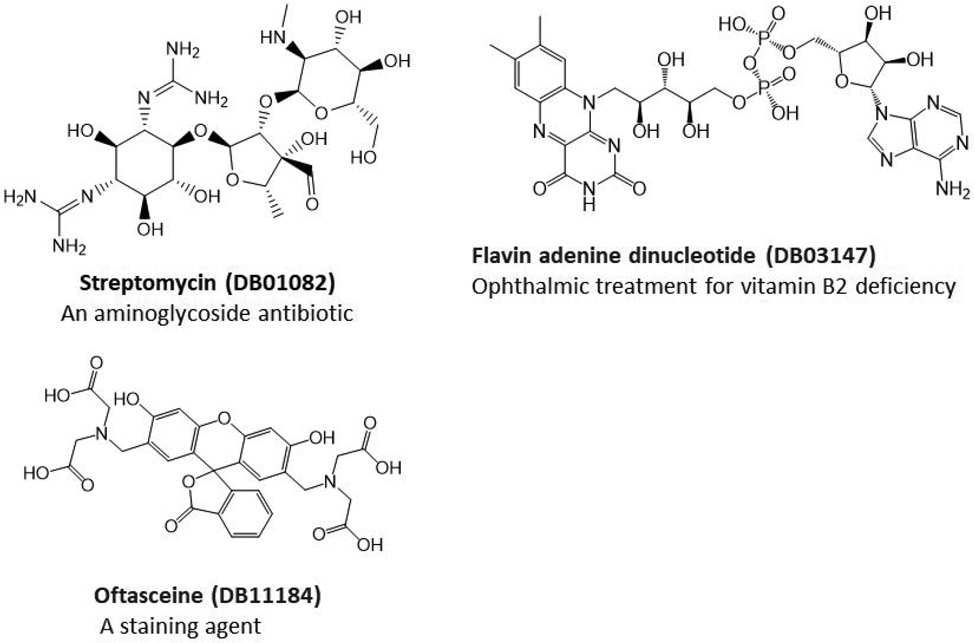
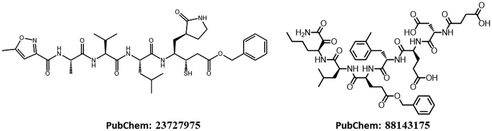
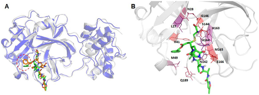
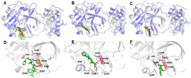
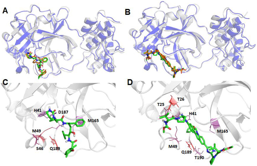
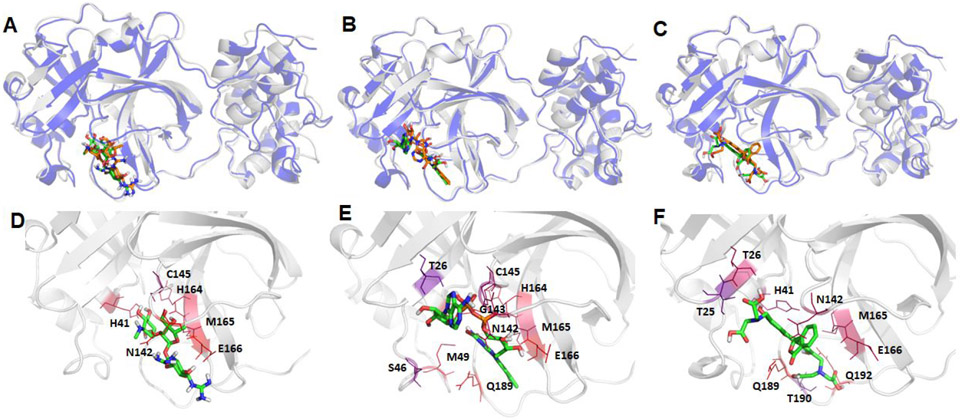
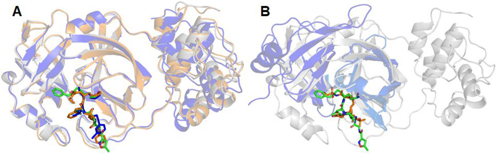
The recent outbreak of novel coronavirus disease-19 (COVID-19) calls for and welcomes possible treatment strategies using drugs on the market. It is very efficient to apply computer-aided drug design techniques to quickly identify promising drug repurposing candidates, especially after the detailed 3D structures of key viral proteins are resolved. The virus causing COVID-19 is SARS-CoV-2. Taking advantage of a recently released crystal structure of SARS-CoV-2 main protease in complex with a covalently bonded inhibitor, N3 (Liu et al., 10.2210/pdb6LU7/pdb), I conducted virtual docking screening of approved drugs and drug candidates in clinical trials. For the top docking hits, I then performed molecular dynamics simulations followed by binding free energy calculations using an end point method called MM-PBSA-WSAS (molecular mechanics/Poisson-Boltzmann surface area/weighted solvent-accessible surface area; Wang, Chem. Rev. 2019, 119, 9478; Wang, Curr. Comput.-Aided Drug Des. 2006, 2, 287; Wang; ; Hou J. Chem. Inf. Model., 2012, 52, 1199). Several promising known drugs stand out as potential inhibitors of SARS-CoV-2 main protease, including carfilzomib, eravacycline, valrubicin, lopinavir, and elbasvir. Carfilzomib, an approved anticancer drug acting as a proteasome inhibitor, has the best MM-PBSA-WSAS binding free energy, -13.8 kcal/mol. The second-best repurposing drug candidate, eravacycline, is synthetic halogenated tetracycline class antibiotic. Streptomycin, another antibiotic and a charged molecule, also demonstrates some inhibitory effect, even though the predicted binding free energy of the charged form (-3.8 kcal/mol) is not nearly as low as that of the neutral form (-7.9 kcal/mol). One bioactive, PubChem 23727975, has a binding free energy of -12.9 kcal/mol. Detailed receptor-ligand interactions were analyzed and hot spots for the receptor-ligand binding were identified. I found that one hot spot residue, His41, is a conserved residue across many viruses including SARS-CoV, SARS-CoV-2, MERS-CoV, and hepatitis C virus (HCV). The findings of this study can facilitate rational drug design targeting the SARS-CoV-2 main protease.


Figures

Figure 1.
Figure 1.
2D-Structures of promising repurpose drugs. All five approved drugs are in neural form under physiological conditions.
Figure 2.
Figure 2.
2D-Structures of promising repurposing drugs. All three approved drugs are in charged form under physiological conditions.
Figure 3.
Figure 3.
2D-Structures of promising bio-actives which are structurally similar to Lopinavir. PubChem 88143175, although studied in neutral form, bears −3 charges under physiological conditions.
Figure 4.
Figure 4.
Plots of Root-mean-square deviations of receptor main chain atoms and ligand heavy atoms along the MD simulation time for (A) co-crystal ligand N3, (B) DB08889, (C) DB12329, (D) DB00385, (E) DB01601 and (F) DB11574.
Figure 5.
Figure 5.
Structural comparison between the crystal structure and a representative MD structure of SARS-Cov-2 main protease bound to the known ligand, N3. The crystal structure is shown as blue cartoon with the co-crystal ligand shown as brown sticks, while the representative MD structure is shown in grey cartoon and the ligand as green sticks (Panel A). The hotspot residues (ΔGLig-Res < −3.0 kcal/mol) revealed by MM-GBSA analysis are shown in Panel B; the more bluish a residue is colored, the stronger the interaction between the residue and the ligand.
Figure 6.
Figure 6.
Structural comparison between the crystal structure and a representative MD structure of SARS-Cov-2 main protease bound to three neutral ligands DB08889, DB12329, and DB00385. The crystal structure is shown as blue cartoon with the docked ligand shown as brown sticks, while the representative MD structure is shown in grey cartoon and the ligand as green sticks. A: DB08889, B: DB12329, and C: DB00385. The detailed ligand-receptor interactions are shown in the bottom panel (D-F). All the hotspot residues (ΔGLig-Res < −3.0) revealed by MM-GBSA analyses are labeled and colored by a blue to red spectrum, the more bluish a residue is colored, the stronger the interaction between the residue and the ligand. D: DB08889, E: DB12329, and F: DB00385.
Figure 7.
Figure 7.
Structural comparison between the crystal and a representative MD structure of SARS-Cov-2 main protease bound to two neutral ligands DB01601 and DB11574. The crystal structure is shown as blue cartoon with the docked ligand shown as brown sticks, while the representative MD structure is shown in grey cartoon and the ligand as green sticks. A: DB01601 and B: DB11574. The detailed ligand-receptor interactions are shown in the bottom panel (C-D). All the hotspot residues (ΔGLig-Res < −3.0) revealed by MM-GBSA analyses are labeled and colored by a blue to red spectrum, the more bluish a residue is colored, the stronger interaction between the residue and the ligand. C: DB01601 and D: DB11574.
Figure 8.
Figure 8.
Structural comparison between the crystal structure and a representative MD structure of SARS-Cov-2 main protease bound to three charged ligands DB01082, DB03147, and DB11184. The crystal structure is shown as blue cartoon with the docked ligand shown as brown sticks, while the representative MD structure is shown in grey cartoon and the ligand as green sticks. A: DB01082, B: DB03147, and C: DB11184. The detailed ligand-receptor interactions are shown in the bottom panel (D-F). All the hotspot residues (ΔGLig-Res < −3.0) revealed by MM-GBSA analyses are labeled and colored by a blue to red spectrum, the more bluish a residue is colored, the stronger the interaction between the residue and the ligand. D: DB01082, E: DB03147, and F: DB11184.
Figure 9.
Figure 9.
Structural comparison of proteases among three coronavirus viruses (SARS-Cov-2, SARS-Cov and MERS-Cov) (A), and between SARS-Cov-2 and hepatitis C NS3/4A proteases (B). The SARS-Cov-2 main protease is colored in grey and its ligands are shown as green sticks. The following are the color codes for the other proteases: SARS-Cov protease and its co-crystal ligand – brown, MERS-Cov protease and its co-crystal ligand – blue, HCV NS3/4A – blue, co-crystal ligand of HCV NS3/4A – brown. Backbone RMSD between SARS-Cov and SARS-Cov-2 is 0.4711 Å, with 284 residues participating in the least-square fitting and 22 omitted, and the backbone RMSD between MERS-Cov and SARS-Cov-2 is 0.41 Å, but with 195 residues participating in the least-square fitting and 104 omitted. In contrast, the backbone RMSD between SARS-Cov-2 main protease and HCV NS3/4A is 2.2632 Å, with 108 residues participating in the least-square fitting and 43 omitted.

Update of

Comment in

  • Cancer Labs Pivot to Battle COVID-19.
    [No authors listed] [No authors listed] Cancer Discov. 2020 May;10(5):634. doi: 10.1158/2159-8290.CD-ND2020-006. Epub 2020 Mar 31. Cancer Discov. 2020. PMID: 32234716

References

    1. Liu X; Zhang B; Jin Z; Yang H; Rao Z, The crystal structure of COVID-19 main protease in complex with an inhibitor N3. 2020.
    1. Wang E; Sun H; Wang J; Wang Z; Liu H; Zhang JZH; Hou T, End-Point Binding Free Energy Calculation with MM/PBSA and MM/GBSA: Strategies and Applications in Drug Design. Chem. Rev 2019, 119, 9478–9508. - PubMed
    1. Wang JM; Hou TJ; Xu XJ, Recent Advances in Free Energy Calculations with a Combination of Molecular Mechanics and Continuum Models. Curr. Comput.-Aided Drug Des 2006, 2, 287–306.
    1. Wang J; Hou T, Develop and test a solvent accessible surface area-based model in conformational entropy calculations. J. Chem. Info. Model 2012, 52, 1199–1212. - PMC - PubMed
    1. Law V; Knox C; Djoumbou Y; Jewison T; Guo AC; Liu Y; Maciejewski A; Arndt D; Wilson M; Neveu V; Tang A; Gabriel G; Ly C; Adamjee S; Dame ZT; Han B; Zhou Y; Wishart DS, DrugBank 4.0: shedding new light on drug metabolism. Nucleic Acids Res. 2014, 42 (Database issue), D1091–1097. - PMC - PubMed

Publication types

MeSH terms