. 2020 Apr 17;10(4):623.
doi: 10.3390/biom10040623.

Physicochemical Characterisation of KEIF-The Intrinsically Disordered N-Terminal Region of Magnesium Transporter A

Affiliations

Physicochemical Characterisation of KEIF-The Intrinsically Disordered N-Terminal Region of Magnesium Transporter A

Stéphanie Jephthah et al. Biomolecules. .

Abstract

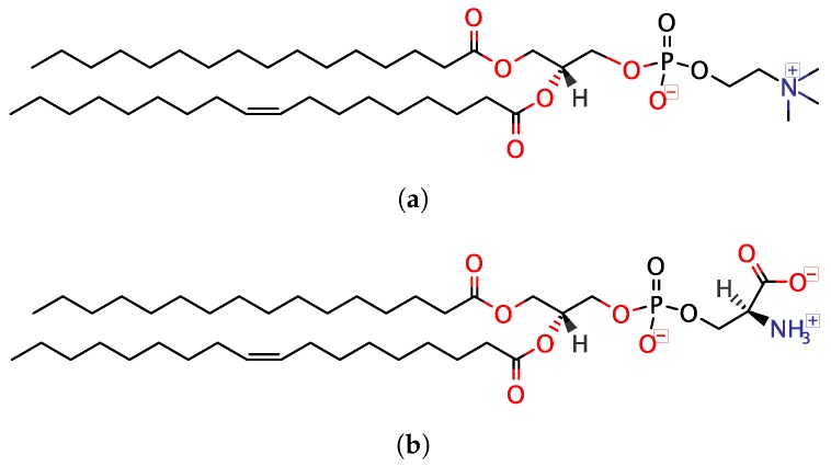
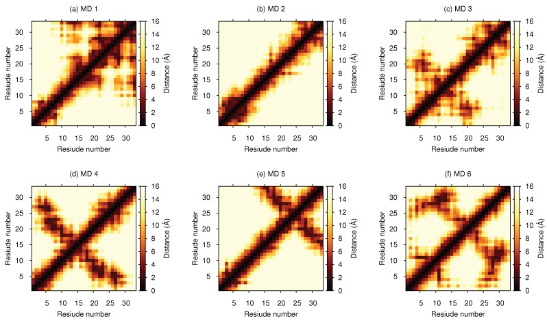
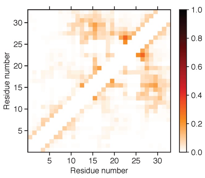
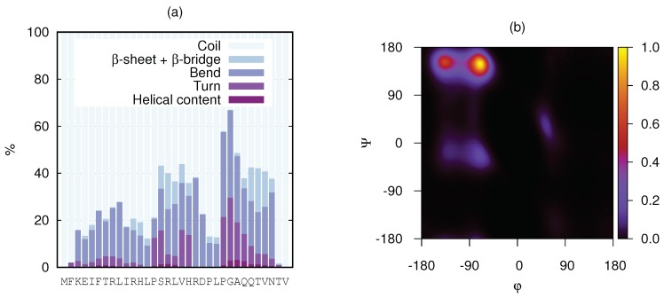
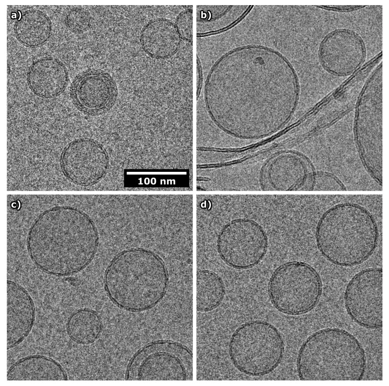
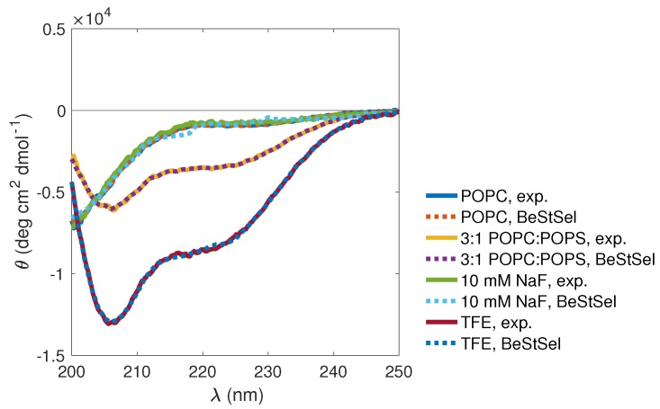
Magnesium transporter A (MgtA) is an active transporter responsible for importing magnesium ions into the cytoplasm of prokaryotic cells. This study focuses on the peptide corresponding to the intrinsically disordered N-terminal region of MgtA, referred to as KEIF. Primary-structure and bioinformatic analyses were performed, followed by studies of the undisturbed single chain using a combination of techniques including small-angle X-ray scattering, circular dichroism spectroscopy, and atomistic molecular-dynamics simulations. Moreover, interactions with large unilamellar vesicles were investigated by using dynamic light scattering, laser Doppler velocimetry, cryogenic transmission electron microscopy, and circular dichroism spectroscopy. KEIF was confirmed to be intrinsically disordered in aqueous solution, although extended and containing little β -structure and possibly PPII structure. An increase of helical content was observed in organic solvent, and a similar effect was also seen in aqueous solution containing anionic vesicles. Interactions of cationic KEIF with anionic vesicles led to the hypothesis that KEIF adsorbs to the vesicle surface through electrostatic and entropic driving forces. Considering this, there is a possibility that the biological role of KEIF is to anchor MgtA in the cell membrane, although further investigation is needed to confirm this hypothesis.

Keywords: circular dichroism spectroscopy; cryogenic transmission electron microscopy; intrinsically disordered proteins; magnesium transporter; membrane proteins; molecular-dynamics simulations; protein–vesicle interactions; secondary structure.; small-angle X-ray scattering.


Conflict of interest statement

The authors declare no conflicts of interest.

Figures

Figure 1
Figure 1
Lipids used for preparation of large unilamellar vesicles (LUVs): (a) 1-palmitoyl-2-oleoyl-glycero-3-phosphocholine (POPC) and (b) 1-palmitoyl-2-oleoyl-sn-glycero-3- phospho-L-serine (POPS).
Figure 2
Figure 2
Primary structure analyses of KEIF: (a) Estimated charge per amino acid residue. Charge of peptide termini included as separate residues and marked as dots in the x-axis label. (b) Das–Pappu plot [33,34]. KEIF location is indicated by white circle in Region R1. (c) Disorder propensity per amino acid (CDisProtCPDB)/CPDB, as described by Uversky (2013) [35]. (d) Probability prediction of disordered regions and disordered binding regions using PrDOS (green) [36], IUPred2A (light purple), and ANCHOR2 (dark purple) algorithms [37]. (e) Whole-residue Wimley–White hydrophobicity indices [14,15] per amino acid residue. (f) Kyte–Doolittle [38] smoothed (five amino acid sliding-window) hydropathy plot based on whole-residue Wimley–White indices.
Figure 3
Figure 3
KEIF circular-dichroism (CD) spectra (solid lines) with BeStSel [5,6] fits (dashed lines), showing the effect of (a) varying salt (NaF) concentration (in TRIS buffer), (b) introducing various divalent cations (10 mM NaF in TRIS buffer), and (c) switching to organic solvent (TFE).
Figure 4
Figure 4
Experiment small-angle X-ray scattering (SAXS) results (grey) compared to ensemble-optimisation-method (EOM; blue) and molecular-dynamics (MD) simulations (red). (a) Form factors, (b) Kratky plot, and (c) distance-distribution functions.
Figure 5
Figure 5
Distance maps depicting distance between amino acid residues for each representative structure of top six clusters from MD simulation.
Figure 6
Figure 6
Illustration of cation–π interaction between Phe-6 and Arg-20 in MD 3 structure. marked distances given in Ångström (Å).
Figure 7
Figure 7
Contact map showing the probability of amino acid residues being closer to each other than cutoff of 4.0 Å. Darker colour indicates higher probability, and white corresponds to zero probability. Residue interactions with themselves, as well as two neighbouring residues on each side, were excluded from analysis.
Figure 8
Figure 8
(a) Stacked secondary-structure histograms per amino acid residue of KEIF as obtained from MD simulations. Three different types of helices included in helical content: (i) α-helix, (ii) 310-helix, and (iii) π-helix. This analysis did not include the PPII helix. (b) Ramachandran plot of KEIF as obtained from MD simulations.
Figure 9
Figure 9
Representative cryo-TEM images of (a,b) POPC and (c,d) 3:1 POPC:POPS vesicles in the (a,c) absence and (b,d) presence of KEIF, at 10 mM NaF in TRIS buffer. The lipid:KEIF molar ratio was 16:1. The scale bar applies to all images.
Figure 10
Figure 10
KEIF CD spectra recorded in presence of POPC and 3:1 POPC:POPS vesicles, in TRIS buffer supplemented with 10 mM NaF. Lipid:KEIF molar ratio was 16:1. For comparison, spectra recorded in TRIS buffer supplemented with 10 mM NaF and in TFE are also shown. Dashed lines represent BeStSel [5,6] fits.
Figure 11
Figure 11
Schematic cross-section of an MgtA-carrying cell membrane. It is possible that KEIF plays the role of an anchor, helping to stabilise the large protein complex in the membrane by locking it in place via electrostatic interactions with anionic lipid head groups.

References

    1. Reinhart R.A. Magnesium metabolism: A review with special reference to the relationship between intracellular content and serum levels. Arch. Intern. Med. 1988;148:2415–2420. doi: 10.1001/archinte.1988.00380110065013. - DOI - PubMed
    1. Maguire M.E. Magnesium transporters: Properties, regulation and structure. Front. Biosci. 2006;11:3149–3163. doi: 10.2741/2039. - DOI - PubMed
    1. Subramani S., Perdreau-Dahl H., Morth J.P. The magnesium transporter A is activated by cardiolipin and is highly sensitive to free magnesium in vitro. eLife. 2016;5:e11407. doi: 10.7554/eLife.11407. - DOI - PMC - PubMed
    1. Jones D.T., Cozzetto D. DISOPRED3: Precise disordered region predictions with annotated protein-binding activity. Bioinformatics. 2015;31:857–863. doi: 10.1093/bioinformatics/btu744. - DOI - PMC - PubMed
    1. Micsonai A., Wien F., Kernya L., Lee Y.H., Goto Y., Réfrégiers M., Kardos J. Accurate secondary structure prediction and fold recognition for circular dichroism spectroscopy. Proc. Natl. Acad. Sci. USA. 2015;112:E3095–E3103. doi: 10.1073/pnas.1500851112. - DOI - PMC - PubMed

Publication types

MeSH terms

LinkOut - more resources