Review
. 2020 Apr 17;10(4):624.
doi: 10.3390/biom10040624.

Structural Insight into the Mechanism of N-Linked Glycosylation by Oligosaccharyltransferase

Affiliations
Review

Structural Insight into the Mechanism of N-Linked Glycosylation by Oligosaccharyltransferase

Smita Mohanty et al. Biomolecules. .

Abstract

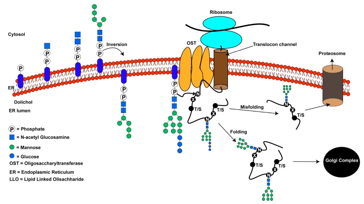
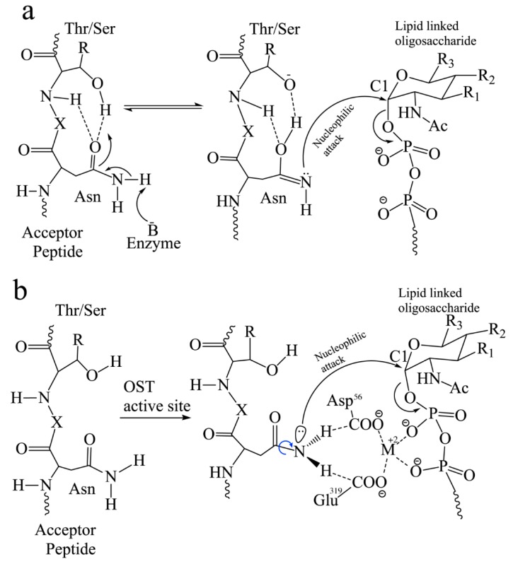
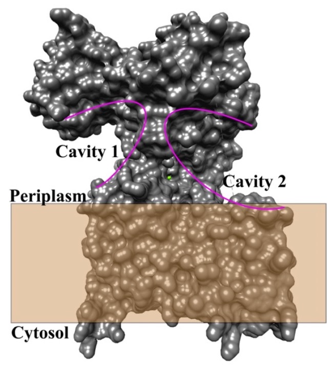
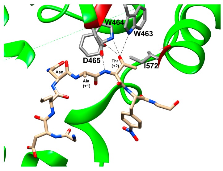
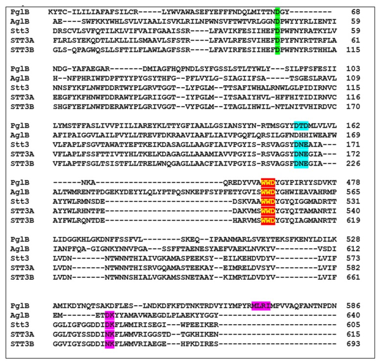
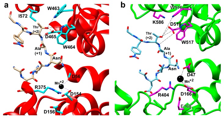
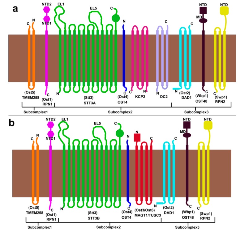
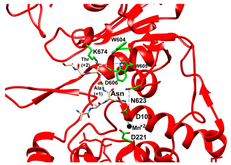
Asparagine-linked glycosylation, also known as N-linked glycosylation is an essential and highly conserved post-translational protein modification that occurs in all three domains of life. This modification is essential for specific molecular recognition, protein folding, sorting in the endoplasmic reticulum, cell-cell communication, and stability. Defects in N-linked glycosylation results in a class of inherited diseases known as congenital disorders of glycosylation (CDG). N-linked glycosylation occurs in the endoplasmic reticulum (ER) lumen by a membrane associated enzyme complex called the oligosaccharyltransferase (OST). In the central step of this reaction, an oligosaccharide group is transferred from a lipid-linked dolichol pyrophosphate donor to the acceptor substrate, the side chain of a specific asparagine residue of a newly synthesized protein. The prokaryotic OST enzyme consists of a single polypeptide chain, also known as single subunit OST or ssOST. In contrast, the eukaryotic OST is a complex of multiple non-identical subunits. In this review, we will discuss the biochemical and structural characterization of the prokaryotic, yeast, and mammalian OST enzymes. This review explains the most recent high-resolution structures of OST determined thus far and the mechanistic implication of N-linked glycosylation throughout all domains of life. It has been shown that the ssOST enzyme, AglB protein of the archaeon Archaeoglobus fulgidus, and the PglB protein of the bacterium Campylobactor lari are structurally and functionally similar to the catalytic Stt3 subunit of the eukaryotic OST enzyme complex. Yeast OST enzyme complex contains a single Stt3 subunit, whereas the human OST complex is formed with either STT3A or STT3B, two paralogues of Stt3. Both human OST complexes, OST-A (with STT3A) and OST-B (containing STT3B), are involved in the N-linked glycosylation of proteins in the ER. The cryo-EM structures of both human OST-A and OST-B complexes were reported recently. An acceptor peptide and a donor substrate (dolichylphosphate) were observed to be bound to the OST-B complex whereas only dolichylphosphate was bound to the OST-A complex suggesting disparate affinities of two OST complexes for the acceptor substrates. However, we still lack an understanding of the independent role of each eukaryotic OST subunit in N-linked glycosylation or in the stabilization of the enzyme complex. Discerning the role of each subunit through structure and function studies will potentially reveal the mechanistic details of N-linked glycosylation in higher organisms. Thus, getting an insight into the requirement of multiple non-identical subunits in the N-linked glycosylation process in eukaryotes poses an important future goal.

Keywords: congenital disorders of glycosylation; cryo-EM structures; human oligosaccharyltransferase; mechanism of N-linked glycosylation; membrane proteins; yeast oligosaccharyltransferase.


Conflict of interest statement

The authors declare no conflict of interest.

Figures

Figure 1
Figure 1
An overview of the N-linked glycosylation reaction of proteins in higher eukaryotes: pyrophosphate and monosaccharides are added to the dolichol lipid on the cytosolic side of the endoplasmic reticulum. The lipid linked oligosaccharide (LLO) is inverted to the luminal side of the endoplasmic reticulum (ER). Additional monosaccharides are added to form the mature LLO. Oligosaccharyltransferase (OST) catalyzes the transfer of the oligosaccharide from the LLO to the side-chain of an asparagine residue in -N-X-T/S- consensus sequence within a protein. Protein folding occurs after N-linked glycosylation. The three terminal glucose residues are trimmed before translocating to the Golgi apparatus for sorting. Misfolded proteins are targeted for degradation by proteasomes.
Figure 2
Figure 2
Possible reaction schemes of N-linked glycosylation showing nucleophilic attack by sidechain amide of the acceptor asparagine residue yielding a glycosylated peptide. (a) Mechanism of formation of an imidate tautomer, a competent nucleophile followed by nucleophilic attack on C1 of the dolichol-linked oligosaccharide. (b) Twisted amide activation mechanism for glycosylation of the acceptor peptide. The amide group forms H- bonds (dashed lines) with Glu319 and Asp56 residues leading to rotation of the C-N bond (indicated by the blue arrow) in bacterial PglB. These residues (Asp56 and Glu319) form H-bonds with the catalytic divalent metal ion. R1 is OH in eukaryotes, and oligosaccharyl in bacteria. R2 is oligosaccharyl in eukaryotes and NHAc in bacteria. R3 is CH2OH in eukaryotes and CH3 in bacteria [58]. X is any amino acid except proline.
Figure 3
Figure 3
Surface representation of the bacterial PglB protein displays two cavities right above the membrane. The cavities are highlighted by purple solid arcs. The figure was prepared with chimera software and PDB file 3RCE.
Figure 4
Figure 4
Residues interacting with the +2 Thr of bound peptide are shown and labelled. Hydrogen bonds from the WWD motif to the β-hydroxyl group are indicated by dashed lines. The figure was prepared using chimera and PDB ID 3RCE [33].
Figure 5
Figure 5
Sequence alignment of bacterial PglB, archaeon AglB, yeast Stt3, human STT3A, and human STT3B proteins to show the important residues and motifs. D56 (PglB), D47 (AglB and yeast Stt3), D49 (human STT3A), and D103 (human STT3B) are shown in green background. DXD motif in PglB, DXE motifs in yeast Stt3, human STT3A, and human STT3B are shown in cyan background. The conserved WWD motif is shown in red background highlighted in yellow. The MXXI motif in PglB that corresponds to DK motifs in AglB and yeast Stt3 are shown in purple background.
Figure 6
Figure 6
Active site of (a) bacterial PglB (PDB ID: 3RCE) and (b) yeast Stt3 (PDB ID: 6EZN), indicating the important residues involved in acceptor peptide recognition for glycosylation and metal ion co-ordination. Residues to metal co-ordination and H- bond of the WWD motif to +2 Thr of the acceptor peptide are shown with dotted lines.
Figure 7
Figure 7
Subunit organization of the metazoan and yeast OST complex in ER membrane. (a) OST-A complex. (b) OST-B complex. Subunits are labeled by mammalian names with yeast subunit names shown in parentheses. Mammalian OST-A complex is homologous to the yeast OST complex, while the yeast OST lacks KCP2 and DC2 subunits found exclusively in the OST-B complex.
Figure 8
Figure 8
Close-up view of structure of STT3B (PDB ID: 6S7T) in cartoon representation. Residues interacting with Thr at +2 position of the acceptor peptide and with metal ion are shown as sticks and labeled. The H-bond formed by WWD motif to +2 Thr and metal to residue co-ordination are shown with dashed lines. D103 and N623 in STT3B correspond to D56 and E319 in bacterial PglB.

References

    1. Dempski R.E., Jr., Imperiali B. Oligosaccharyl transferase: Gatekeeper to the secretory pathway. Curr. Opin. Chem. Biol. 2002;6:844–850. doi: 10.1016/S1367-5931(02)00390-3. - DOI - PubMed
    1. Kornfeld R., Kornfeld S. Assembly of asparagine-linked oligosaccharides. Annu. Rev. Biochem. 1985;54:631–664. doi: 10.1146/annurev.bi.54.070185.003215. - DOI - PubMed
    1. Larkin A., Imperiali B. The expanding horizons of asparagine-linked glycosylation. Biochemistry. 2011;50:4411–4426. doi: 10.1021/bi200346n. - DOI - PMC - PubMed
    1. Wacker M., Linton D., Hitchen P.G., Nita-Lazar M., Haslam S.M., North S.J., Panico M., Morris H.R., Dell A., Wren B.W., et al. N-linked glycosylation in Campylobacter jejuni and its functional transfer into E. coli. Science. 2002;298:1790–1793. doi: 10.1126/science.298.5599.1790. - DOI - PubMed
    1. Mohorko E., Glockshuber R., Aebi M. Oligosaccharyltransferase: The central enzyme of N-linked protein glycosylation. J. Inherit. Metab. Dis. 2011;34:869–878. doi: 10.1007/s10545-011-9337-1. - DOI - PubMed

Publication types

Substances

LinkOut - more resources