Review
. 2020 Apr 21;24(1):163.
doi: 10.1186/s13054-020-02885-8.

Brain-heart interaction after acute ischemic stroke

Affiliations
Review

Brain-heart interaction after acute ischemic stroke

Denise Battaglini et al. Crit Care. .

Abstract

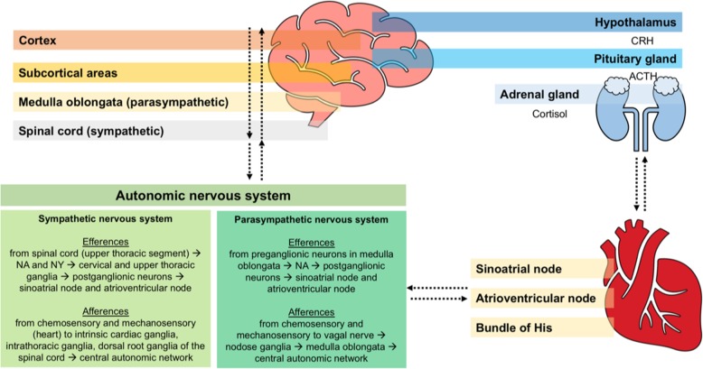
Early detection of cardiovascular dysfunctions directly caused by acute ischemic stroke (AIS) has become paramount. Researchers now generally agree on the existence of a bidirectional interaction between the brain and the heart. In support of this theory, AIS patients are extremely vulnerable to severe cardiac complications. Sympathetic hyperactivity, hypothalamic-pituitary-adrenal axis, the immune and inflammatory responses, and gut dysbiosis have been identified as the main pathological mechanisms involved in brain-heart axis dysregulation after AIS. Moreover, evidence has confirmed that the main causes of mortality after AIS include heart attack, congestive heart failure, hemodynamic instability, left ventricular systolic dysfunction, diastolic dysfunction, arrhythmias, electrocardiographic anomalies, and cardiac arrest, all of which are more or less associated with poor outcomes and death. Therefore, intensive care unit admission with continuous hemodynamic monitoring has been proposed as the standard of care for AIS patients at high risk for developing cardiovascular complications. Recent trials have also investigated possible therapies to prevent secondary cardiovascular accidents after AIS. Labetalol, nicardipine, and nitroprusside have been recommended for the control of hypertension during AIS, while beta blockers have been suggested both for preventing chronic remodeling and for treating arrhythmias. Additionally, electrolytic imbalances should be considered, and abnormal rhythms must be treated. Nevertheless, therapeutic targets remain challenging, and further investigations might be essential to complete this complex multi-disciplinary puzzle. This review aims to highlight the pathophysiological mechanisms implicated in the interaction between the brain and the heart and their clinical consequences in AIS patients, as well as to provide specific recommendations for cardiovascular management after AIS.

Keywords: Acute ischemic stroke; Arrhythmia; Cardiovascular; Cerebrovascular; Heart; Neuroinflammation.


Conflict of interest statement

The authors declare that they have no competing interests.

Figures

Fig. 1
Fig. 1
Neural connections: from the brain to the heart and from the heart to the brain. Sympathetic system efferences from the spinal cord (NA and NY) to the cervical and upper thoracic ganglia, thus to sinus atrial node and atrial ventricular node. Afferences from the chemosensory and mechanosensory neurons in the heart to the intrathoracic and dorsal root ganglia of the spinal cord, to central autonomic network. Parasympathetic system efferences from the medulla oblongata (noradrenaline) to the sinus atrial node and atrial ventricular node. Afferences from the chemosensory and mechanosensory neurons to the vagal nerve, nodose ganglia, medulla oblongata, and central autonomic network. Efferences from the hypothalamus (CRH) to the pituitary gland (ACTH) to the adrenal gland (cortisol). NA, noradrenaline; NY, neuropeptide Y; CRH, corticotropin-releasing hormone; ACTH, adrenocorticotropic hormone
Fig. 2
Fig. 2
Activation of different brain areas during stroke followed by specific cardiovascular complications. Depending on the extent of subsequent brain damage, stroke triggers different central regulatory regions, thus activating corresponding pathways that depend on the injured area. Therefore, post-stroke cardiac dysfunctions may be referred to specific brain areas. Right-sided stroke is usually associated with more cardiac complications than left-sided stroke. QTc, corrected QT interval
Fig. 3
Fig. 3
Brain–heart sympathetic pathway at the molecular level. The “fight or flight” response of catecholaminergic storm, followed by hypothalamic–pituitary–adrenal axis and autonomic activation, is represented at the molecular level. Synaptic connection through neurons and myocytes is represented. Noradrenaline activates β1 receptors, which in turn activates cyclic adenosine monophosphate–protein kinase A (cAMP–PKA) signaling, with consequent release of Ca2+ from the sarcoplasmic reticulum for cell contraction. At the same time, noradrenaline activates β2 receptors, which, acting through the protein kinase B (Akt)-FOXO pathway, decrease protein degradation by ubiquitin, thus regulating cardiomyocyte proteostatic equilibrium and cardiac mass maintenance with muscle ring finger-1 (MuRF-1), which is upregulated in the deficient heart. FOXO, forkhead box O; Akt, protein kinase B; PKA, protein kinase A; cAMP, cyclic adenosine monophosphate, ATP, adenosine triphosphate; MuRF-1, muscle ring finger-1. Modified from "Martini FH. Fundamentals of Anatomy and Physiology. 8th ed. 2006. Chapter 20"
Fig. 4
Fig. 4
Local inflammatory response after stroke. The local inflammatory process starts with the activation of pro-inflammatory and pro-coagulative cascades into the intravascular space. The blood–brain barrier disruption allows the infiltration of peripheral macrophages and neutrophils into the ischemic lesion. This leads to an enhanced local inflammatory response in the brain parenchyma. Other peripheral immune cells are thus recalled into the ischemic brain and cerebral microcirculation, subsequently crossing the damaged blood–brain barrier and passing into the systemic circulation. ROS, reactive oxygen species; TNF-α, tumor necrosis factor alpha; MMPs, matrix metalloproteinases; DAMPs, danger-associated molecular patterns
Fig. 5
Fig. 5
Gut dysbiosis and cardiac dysfunction. Gut dysbiosis causes increased gut–blood barrier permeability and pathogen translocation, with possible atherosclerosis and thrombosis. Gut pathogens contribute to enhance the inflammatory response through platelet hyperactivation and thrombosis, mediated by the conversion of choline and l-carnitine into trimethylamine N-oxide (TMAO). TMAO induces platelet hyperactivity and foam cell formation, alters bile and sterol metabolism, and activates macrophages, dendritic cells, and platelets. TMAO, trimethylamine N-oxide
Fig. 6
Fig. 6
Flow chart of stroke management after intensive care unit admission. Summary of the first steps for the detection and management of possible cardiac complications in stroke patients after intensive care unit admission. Due to the lack of conclusive data, no specific recommendations on pharmacotherapy are given; readers are strongly advised to follow local protocols and international guidelines. ECG, electrocardiogram; BP, blood pressure; ICU, intensive care unit; CPR, cardiopulmonary resuscitation

References

    1. Wolf PA, Abbott RD, Kannel WB. Atrial fibrillation as an independent risk factor for stroke: the Framingham study. Stroke. 1991;22:983–988. doi: 10.1161/01.STR.22.8.983. - DOI - PubMed
    1. Chen Z, Venkat P, Seyfried D, Chopp M, Yan T, Chen J. Brain-heart interaction: cardiac complications after stroke. Circ Res. 2017;121:451–468. doi: 10.1161/CIRCRESAHA.117.311170. - DOI - PMC - PubMed
    1. Fuhrer H, Reinhard M, Niesen WD. Paradigm change? Cardiac output better associates with cerebral perfusion than blood pressure in ischemic stroke. Front Neurol. 2017;8:706. - PMC - PubMed
    1. Sposato LA, Cipriano LE, Saposnik G, Vargas ER, Riccio PM, Hachinski V. Diagnosis of atrial fibrillation after stroke and transient ischaemic attack: a systematic review and meta-analysis. Lancet Neurol. 2015;14:377–387. doi: 10.1016/S1474-4422(15)70027-X. - DOI - PubMed
    1. Prosser J, MacGregor L, Lees KR, Diener HC, Hacke W, Davis S. Predictors of early cardiac morbidity and mortality after ischemic stroke. Stroke. 2007;38:2295–2302. doi: 10.1161/STROKEAHA.106.471813. - DOI - PubMed

Publication types