. 2020 Apr 22;5(1):39.
doi: 10.1038/s41392-020-0133-y.

Exosomes with low miR-34c-3p expression promote invasion and migration of non-small cell lung cancer by upregulating integrin α2β1

Affiliations

Exosomes with low miR-34c-3p expression promote invasion and migration of non-small cell lung cancer by upregulating integrin α2β1

Wenjing Huang et al. Signal Transduct Target Ther. .

Abstract

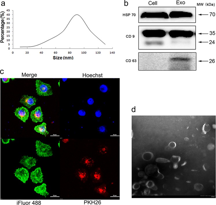
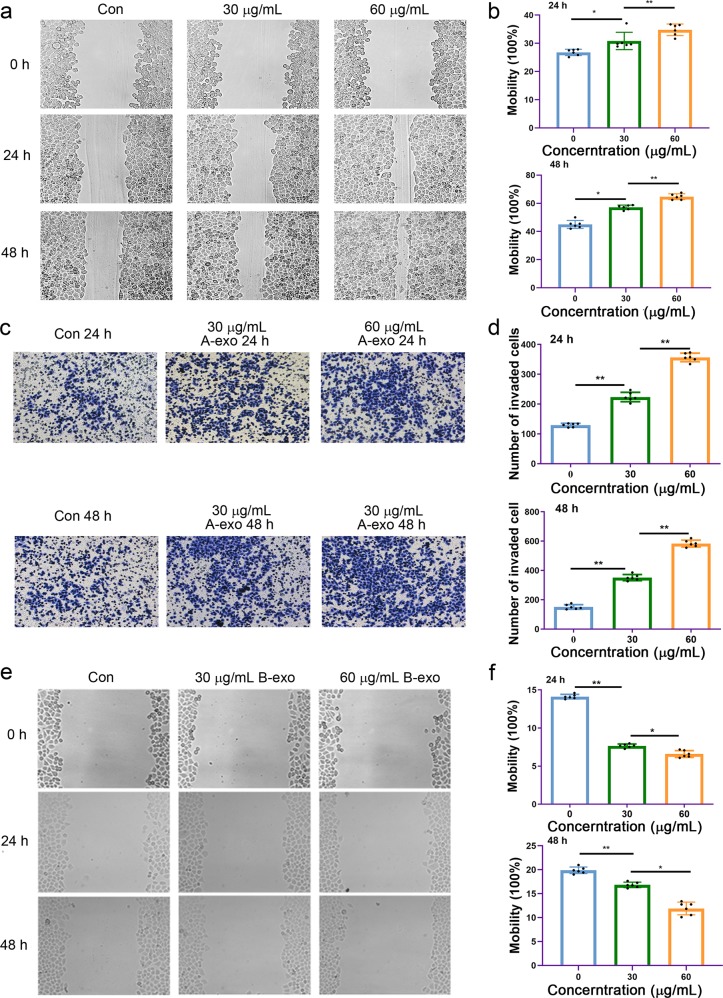
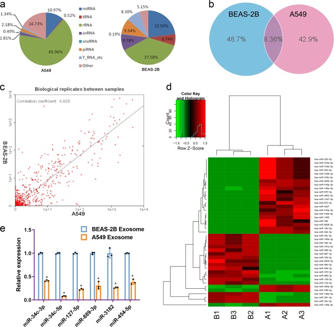
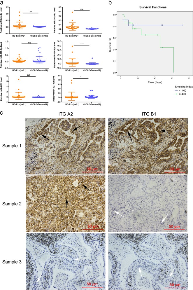
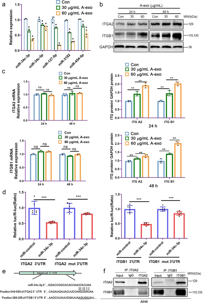
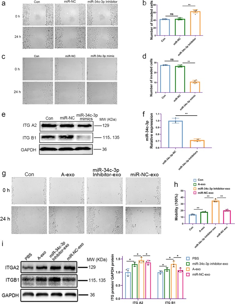
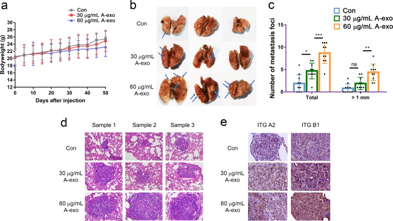
Exosomes play critical roles in regulating various physiological and pathological processes, including immune stimulation, immune suppression, cardiovascular diseases, and cancers. Recent studies show that exosomes that transport specific microRNAs (miRNAs) are involved in tumor development. However, the molecular mechanism by which tumor invasion and migration are regulated by exosomes from non-small cell lung cancer (NSCLC) is not well understood. Here, we show that exosomes shuttling low levels of miR-34c-3p are involved in NSCLC progression. Our results showed that exosomes derived from NSCLC cells carrying low levels of miR-34c-3p could be transported into the cytoplasm of NSCLC cells and accelerate NSCLC invasion and migration by upregulating integrin α2β1. A luciferase assay revealed that integrin α2β1 was the direct target of miR-34c-3p, and overexpression of integrin α2β1 could promote the invasion and migration of NSCLC cells. The analysis of exosomes derived from clinical serum samples indicated that the expression of miR-34c-3p was significantly downregulated in exosomes from NSCLC patients compared with that of normal controls. A549-derived exosomes promoted NSCLC cells lung metastases in vivo. Exosomes shuttling low levels of miR-34c-3p were associated with the progression of NSCLC in vitro and in vivo. Our data demonstrate that exosomes shuttling low levels of miR-34c-3p can accelerate the invasion and migration of NSCLC by upregulating integrin α2β1. MiR-34c-3p can be a diagnostic and prognostic marker for NSCLC. High expression of integrin α2β1 is positively related to the migration and metastasis of NSCLC cells.


Conflict of interest statement

The authors declare no competing interests.

Figures

Fig. 1
Fig. 1
Characterization and uptake of exosomes. a A549-derived exosomes (A-exo) were determined to be between 30 and 120 nm in size by nanoparticle tracking analysis. b Western blots for HSP70, CD9, and CD63 in exosomes and cells. c Confocal microscopy of A549 cells treated with A549 exosomes labeled with the fluorescent linker PKH26 (red). The cytoskeleton of A549 cells was labeled with iFlour 488 Reagent (green), and the nucleus of A549 cells was labeled with Hoechst (blue). d Transmission electron microscopy image of exosomes are shown
Fig. 2
Fig. 2
Increased invasion and migration of A549 cells following treatment with exosomes derived from NSCLC. a NSCLC A549 cell migration was induced by exosomes derived from A549 cells (A-exo) in a dose- and time-dependent manner. b Quantitative results of a. ns no significance, *p < 0.05, **p < 0.01, and n ≥ 3. c A549 cell invasion was induced by A549 cell-derived exosomes (A-exo) in a dose- and time-dependent manner. d Quantitative results of c. *p < 0.05, **p < 0.01, and n ≥ 3. e A549 cell migration was inhibited by BEAS-2B cell-derived exosomes (B-exo) in a dose-dependent manner. f Quantitative results of e. ns no significance, *p < 0.05, **p < 0.01, and n ≥ 3
Fig. 3
Fig. 3
Profiles of miRNA expression in exosomes derived from different cells. a The ratio of the percentage small RNA categories in all reads mapped to noncoding RNA databases. b Venn diagram showing the unique and overlapping miRNAs between exosomes from different cells. c Biological replicates between exosomes from BEAS-2B and A549 cells. d Heatmap diagram of differential miRNA expression between exosomes derived from BEAS-2B cells and A549 cells. Gene expression data were obtained using next-generation sequencing on an Illumina HiSeq 2500 platform. Mean expression values are shown. Red, increased expression; green, decreased expression; and black, mean value. **p < 0.01. e Determination of miR-34c-3p, miR-34c-5p, miR-127-5p, miR-889-3p, miR-3182 and miR-454-5p microRNA levels by quantitative real-time PCR in exosomes from NSCLC cells. Experiments were performed in triplicate
Fig. 4
Fig. 4
Integrin α2β1 protein and exosomal miRNA expression in clinical samples. a Levels of miRNAs in the sera of NSCLC patients and healthy controls. The levels of exosomal miR-34c-3p, miR-34c-5p, miR-889-3p, miR-454-5p, miR-3182, and miR-127-5p in the sera of NSCLC patients (n = 37) and healthy controls (n = 21) were measured by RT-qPCR. Differences in miRNA expression between NSCLC patients and healthy controls were analyzed by nonparametric test, ns no significance, *p < 0.05, **p < 0.01, and ***p < 0.001. b Kaplan-Meier curve analysis of NSCLC patients (n = 37) was performed according to the smoking index. c The protein expression of integrin α2 and β1 in NSCLC tissue samples (n = 50). Immunohistochemistry on the expression of integrin α2 and β1 in 50 human lung adenocarcinoma samples. Three representative staining images are shown, where either integrin α2 or integrin β1 staining was positive (white arrow) or negative (black arrow). ITG A2 Integrin α2, and ITG B1 Integrin β1
Fig. 5
Fig. 5
NSCLC exosomes increased the protein levels of integrin α2β1 by reducing miR-34c-3p. a NSCLC exosomes reduced the expression of miR-34c-3p. Cellular miR-34c-3p levels were detected by quantitative RT-PCR after treatment with NSCLC exosomes. b NSCLC exosomes increased the protein levels of integrin α2β1 in NSCLC cells. Western blotting was performed with the indicated antibodies, and GAPDH was used as a control to normalize levels. c NSCLC exosomes did not affect the mRNA levels of integrin α2β1 in NSCLC cells. The mRNA levels of integrin α2β1 in cells were detected by quantitative RT-PCR after treatment with NSCLC exosomes. d Luciferase activity by luciferase reporters carrying the 3′-UTR of ITGA2 or ITGB1 or mutant ITGA2 or ITGB1 in which the binding site for miR-34c-3p was mutated. The vectors were introduced into 293 T cells along with a negative miR-control (NC) or miR-34c-3p. e Alignment of ITGA2 and ITGB1 3′-UTRs with miR-34c-3p. f The results of coimmunoprecipitation reflect the fact that integrin α2 interacts with integrin β1 in NSCLC cells. ns no significance, *p < 0.05, **p < 0.01, ***p < 0.001, and n ≥ 3. ITG A2 Integrin α2, and ITG B1 Integrin β1
Fig. 6
Fig. 6
Inhibition of miR-34c-3p in exosomes induced A549 cell migration by regulating integrin α2β1. a Migration of A549 cells was induced by transfection with miR-34c-3p inhibitor. b Data analysis of a. c A549 cell migration was inhibited by transfection with miR-34c-3p mimics. d Data analysis of c. ns no significance, *p < 0.05, **p < 0.01, and n ≥ 3. e Western blot of integrin α2β1 expression in A549 cells after transfection with miR-34c-3p mimics. f Reduction in the level of miR-34c-3p in exosomes upon transfection with a miR-34c-3p inhibitor in A549 cells. g Induction of A549 cell migration by miR-34c-3p inhibitor-exo. h Data analysis of Fig. 6g. i Western blot analysis of integrin α2β1 expression from different groups, and GAPDH was used as a control to normalize levels. ns no significance, *p < 0.05, **p < 0.01, ***p < 0.001, and n ≥ 3. ITG A2 Integrin α2, and ITG B1 Integrin β1
Fig. 7
Fig. 7
A549-derived exosomes promoted NSCLC cell metastasis in vivo. a A549-derived exosome treatment caused loss of bodyweight in mice. b Exosome-incubated A549 cells developed more or larger metastatic nodules in the lungs than untreated cells, and the effect was dose-dependent manner. c The statistical analysis of total metastatic foci and metastatic foci with diameters greater than 1 mm, ns no significance, *p < 0.05, **p < 0.01, and ***p < 0.001. d H&E staining of tumor specimens showed that the higher concentration of A549 exosomes caused increased malignant metastases (200×). e A549-derived exosomes promoted NSCLC cell metastasis in vivo in a dose-dependent manner, and the highly metastatic samples showed a higher expression of ITGA2 and ITGB1. ITG A2 Integrin α2, and ITG B1 Integrin β1 (400×)

References

    1. Bray F, et al. Global cancer statistics 2018: GLOBOCAN estimates of incidence and mortality worldwide for 36 cancers in 185 countries. Ca. Cancer J. Clin. 2018;68:394–424. doi: 10.3322/caac.21492. - DOI - PubMed
    1. Vyse S, Huang PH. Targeting EGFR exon 20 insertion mutations in non-small cell lung cancer. Signal Transduct. Target. Ther. 2019;4:5. doi: 10.1038/s41392-019-0038-9. - DOI - PMC - PubMed
    1. Mountain CF. Revisions in the international system for staging lung cancer. Chest. 1997;111:1710–1717. doi: 10.1378/chest.111.6.1710. - DOI - PubMed
    1. Jiang W, Cai G, Hu P, Wang Y. Personalized medicine in non-small cell lung cancer: a review from a pharmacogenomics perspective. Acta Pharm. Sin. B. 2018;8:530–538. doi: 10.1016/j.apsb.2018.04.005. - DOI - PMC - PubMed
    1. Wan Z, et al. Exosome-mediated cell-cell communication in tumor progression. Am. J. Cancer Res. 2018;8:1661–1673. - PMC - PubMed

Publication types

MeSH terms