Review
. 2020 Apr 3:11:407.
doi: 10.3389/fphar.2020.00407. eCollection 2020.

Organoids and Microphysiological Systems: New Tools for Ophthalmic Drug Discovery

Affiliations
Review

Organoids and Microphysiological Systems: New Tools for Ophthalmic Drug Discovery

Jing Bai et al. Front Pharmacol. .

Abstract

Organoids are adept at preserving the inherent complexity of a given cellular environment and when integrated with engineered micro-physiological systems (MPS) present distinct advantages for simulating a precisely controlled geometrical, physical, and biochemical micro-environment. This then allows for real-time monitoring of cell-cell interactions. As a result, the two aforementioned technologies hold significant promise and potential in studying ocular physiology and diseases by replicating specific eye tissue microstructures in vitro. This miniaturized review begins with defining the science behind organoids/MPS and subsequently introducing methods for generating organoids and engineering MPS. Furthermore, we will discuss the current state of organoids and MPS models in retina, cornea surrogates, and other ocular tissue, in regards to physiological/disease conditions. Finally, future prospective on organoid/MPS will be covered here. Organoids and MPS technologies closely recapture the in vivo microenvironment and thusly will continue to provide new understandings in organ functions and novel approaches to drug development.

Keywords: 3D tissue constructs; microphysiological system; ocular; organ-on-a-chip; organoid.


Figures

Figure 1
Figure 1
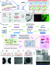
Ocular organoids and microphysiological systems (MPS). (A) Structure of eye, cornea, and retina. Retina structure was adapted from (Fu et al., 2019). (B) Schematic representation on the stepwise generation of organoids. (C) Differentiation of human iPSCs into retinal and corneal organoids. Distinct circular to oval-shaped eye field primordial (EFP) encompassed a centrally located p63+ Recoverin+ neuroretinal (NR) cup (asterisks), adapted from (Susaimanickam et al., 2017). (D) a. Schematic representation of photolithography for polydimethylsiloxane (PDMS)-based microfluidic device fabrication and essential components in a standard MPS model. b.c.d.e.f. An representative example of merging organoid and MPS technology in a human retina-on-a-chip platform: b.c.d. microfluidic platform enabling co-culture of human induced pluripotent stem cell (hiPSC)-derived retinal pigment epithelium (RPE) and ROs (retinal organoids) in a defined physiological structure (the top layer has compartments for the ROs and RPE, and the bottom layer with a channel for a vasculature); e.f. electron microscopic image of polarized RPE cells. RPE cells display apical microvilli (top row) and a basal membrane (bottom row), and immunohistochemical staining for ezrin (green), an apical microvilli marker, indicated polarized RPE, adapted from (Achberger et al., 2019).

References

    1. Achberger K., Probst C., Haderspeck J., Bolz S., Rogal J., Chuchuy J., et al. (2019). Merging organoid and organ-on-a-chip technology to generate complex multi-layer tissue models in a human retina-on-a-chip platform. Elife 8, e46188 . 10.7554/eLife.46188 - DOI - PMC - PubMed
    1. Adriani G., Bai J., Wong S., Kamm R. D., Thiery J. P. (2016). M2a macrophages induce contact-dependent dispersion of carcinoma cell aggregates. Macrophage 3, e1222. 10.14800/Macrophage.1222 - DOI - PMC - PubMed
    1. Aref A. R., Huang R. Y., Yu W., Chua K. N., Sun W., Tu T. Y., et al. (2013). Screening therapeutic EMT blocking agents in a three-dimensional microenvironment. Integr. Biol. (Camb) 5, 381–389. 10.1039/C2IB20209C - DOI - PMC - PubMed
    1. Bai J., Adriani G., Dang T. M., Tu T. Y., Penny H. X., Wong S. C., et al. (2015. a). Contact-dependent carcinoma aggregate dispersion by M2a macrophages via ICAM-1 and beta2 integrin interactions. Oncotarget 6, 25295–25307. 10.18632/oncotarget.4716 - DOI - PMC - PubMed
    1. Bai J., Tu T. Y., Kim C., Thiery J. P., Kamm R. D. (2015. b). Identification of drugs as single agents or in combination to prevent carcinoma dissemination in a microfluidic 3D environment. Oncotarget 6, 36603–36614. 10.18632/oncotarget.5464 - DOI - PMC - PubMed